High-Field MR System Installed at Leading University Medical Center in Holland
By MedImaging International staff writers
Posted on 07 Jun 2016
A leading imaging vendor and a company creating clinical solutions for treating cancer and brain disorders are installing a third high-field 1.5T Magnetic Resonance (MR)-guided linear accelerator at a Dutch university medical center.Posted on 07 Jun 2016
The system is intended for research into how real-time clinical imaging and precision radiotherapy can improve outcomes for patients. The system is designed to provide clinicians with a responsive interventional approach by capturing capture high-quality images of tumors and nearby tissue, and enabling them to assess radiation treatment and modifying it if required.

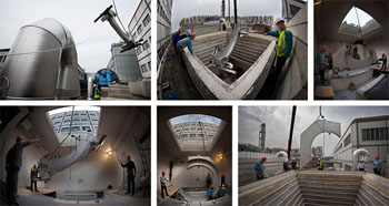
Image: The installation of the high-field MR-guided linear accelerator system at the University Medical Center Utrecht (Photo courtesy of Philips Healthcare).
The installation at the University Medical Center Utrecht (UMC; Utrecht, Netherlands) was announced by Elekta (Stockholm, Sweden) and Philips (Amsterdam, the Netherlands). The Elekta MR-linac system uses a radiotherapy system, a high-field MRI scanner, and software to enable clinicians to view patient anatomy in real time. The MR-linac is designed reduce the radiation exposure of normal tissue, and improve targeting of tumor tissue. The system can locate a tumor and lock onto it during radiotherapy even if the tumor moves or changes shape between treatment sessions. Elekta’s MR-linac is a work in progress and is not available for sale, or distribution.
Bas Raaymakers, PhD, professor at the Department of Radiotherapy, UMC Utrecht, said, “UMC Utrecht has been a leading proponent of the power of MR-linac technology to transform radiotherapy, and we are excited to announce expanded capabilities through installation of a third system. The ability to visualize radiation therapy during treatment and to adapt treatment in real-time based on detailed MR images would allow us to treat cancer with unprecedented levels of precision and accuracy, while improving efficacy and reducing side effects. Just as surgeons require a clear view of the surgical field, and often require sophisticated imaging equipment, radiation oncologists need a 21st century approach to visualize and adapt the radiotherapy field to achieve optimum outcomes for patients.”
Related Links:
University Medical Center Utrecht
Elekta
Philips


