MRI System Helps Plan Prostate Cancer Radiation Treatment
By MedImaging International staff writers
Posted on 11 Apr 2016
An innovative imaging system helps plan radiotherapy (RT) treatment of prostate cancer using magnetic resonance imaging (MRI) alone, without the need for computerized tomography (CT).Posted on 11 Apr 2016
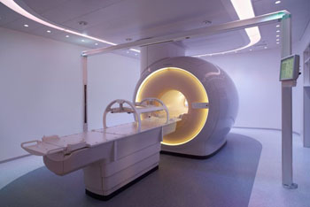
The Royal Philips (Amsterdam, The Netherlands) Magnetic Resonance for Calculating Attenuation (MRCAT) system, designed for the Ingenia MR-RT imaging platform, will support those who choose to use MRI as their single-modality imaging approach to prostate cancer RT treatment planning. The MRCAT system provides high-quality, soft-tissue contrast for target delineation, as well as density information for dose calculations via robust imaging protocols that allow the system to obtain CT-like workflow, and potentially reduced provider costs, as compared to MR-CT workflow.

Image: The Philips Ingenia MR-RT imaging platform (Photo courtesy of Royal Philips).
“Where CT solutions have played a leading role in past radiotherapy treatments, MR takes an innovative approach by providing physicians with increased soft-tissue visualization and functional imaging capabilities to help improve treatment plans,” said Lizette Warner, PhD, manager of clinical science MR therapy at Philips North America. “MR-only simulation makes MR more accessible for hospitals and physicians, transforming the way care is delivered and supporting our customers in improving care for oncology patients who require radiotherapy.”
“Successful cancer treatment depends on the quality and accuracy of the radiation therapy plan, making imaging a critical piece in determining course of treatment. The real power of MR-only simulation is that it enables us to develop personal treatment plans,” said Rodney Ellis, MD, vice chairman of radiation oncology at the Seidman Cancer Center (Cleveland, OH, USA). “The new process will help streamline workflow that will, in turn, reduce the burden on patients and health care providers. Moreover, it can eliminate the systematic errors introduced by MR-CT registration.”
Prostate cancer is the most common cancer among American men, with approximately one million patients undergoing RT annually. Current clinical practice often uses a combined approach of both MR and CT images, but this can lead to image misalignment and registration uncertainties that could impact targeting and treatment. It also puts pressure on patient burden, workflows, and costs.
Related Links:
Royal Philips
Seidman Cancer Center


 Guided Devices.jpg)


