Novel MRI Technique Can Help Diagnose Traumatic Brain Injuries
By MedImaging International staff writers
Posted on 29 Mar 2015
A new, advanced Magnetic Resonance Imaging (MRI) technique that can help detect subtle Traumatic Brain Injuries (TBI) has been developed by the US National Institute of Standards and Technology (NIST; Boulder, CO, USA). Mild TBI is a common injury among war veterans and is difficult to diagnose and treat. TBI can result in symptoms such as memory loss or inability to concentrate.Posted on 29 Mar 2015
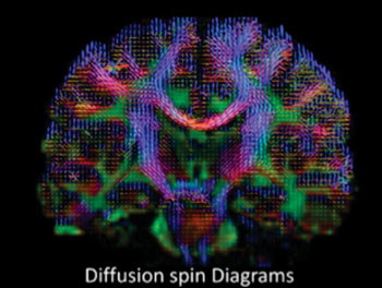
The technique images the brain using High-Definition Fiber Tracking (HDFT) MRI of water diffusion. The NIST creates reference objects called MRI phantoms that enable quantitative measurements to be made according to international standards, and are used to calibrate MRI scanners. The NIST developed an MRI phantom to measure anisotropic diffusion that can show structural information related to TBI such as abnormal neural pathways, nerve cell damage, and the integrity of brain tissue.

Image: High-definition MRI of water diffusion for studies of Traumatic Brain Injury (Photo courtesy of Sudhir Pathak & Walter Schneider/University of Pittsburgh).
The NIST plans to conduct a clinical study of TBI at US Department of Veterans Affairs (VA) hospitals to help reliably diagnose TBI, predict outcomes, and the required care. The research plan, states, “The study is highly relevant to the VA Hospital System commitment to provide a high level of care for veterans with suspected TBI. It will allow a means to optimize scanner performance across the VA system and provide more uniform data from various scanners. Meeting this goal will, for the first time, allow large collections of TBI data to be combined into a single pool. Analysis of the resultant large pool of data is expected to yield important results in terms of early diagnosis of TBI, stratification of patients into treatment categories, assessment of therapeutic results, and data for long-term outcomes trials.”
Related Links:
NIST Boulder


