Characteristic Sign Allows for MR Diagnosis of Parkinson’s Disease
By MedImaging International staff writers
Posted on 12 May 2014
An image similar in shape to the swallow’s tail [a deeply forked tail] has been identified as a new and effective way to diagnose Parkinson’s disease (PD). The image, which depicts the healthy state of a group of cells in a subregion of the human brain, was depicted using 3T magnetic resonance imaging (MRI) scanning technology—conventional equipment currently used in clinical environments.Posted on 12 May 2014
The research was led by Dr. Stefan Schwarz and Prof. Dorothee Auer, experts in neuroradiology in the School of Medicine at the University of Nottingham (UK), and was conducted at the Queen’s Medical Center (Nottingham, UK) in collaboration with Dr. Nin Bajaj, a specialist in movement disorder diseases at the Nottingham University Hospitals National Health Service (NHS) Trust. The findings were been published April 7, 2014, in the open access academic journal PLOS one. The research builds on a successful collaboration with Prof. Penny Gowland at the Sir Peter Mansfield Magnetic Resonance Center at the University of Nottingham.
.")
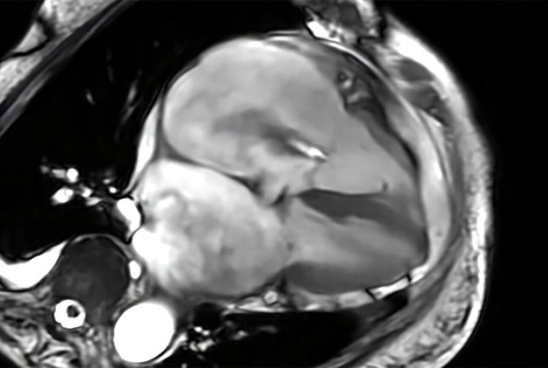
Image: A shape similar to a swallow’s tail has been identified as a new and accurate test for Parkinson’s disease. The image, which depicts the healthy state of a group of cells in the subregion of the human brain, was revealed using 3T MRI scanning technology (Photo courtesy of the University of Nottingham).
The investigators reported that the absence of this imaging sign can help to diagnose PD using conventional clinical MRI scanners. Diagnosing Parkinson’s, up to now, has been clinically uncertain. Diagnosing patients had to rely on expensive nuclear medical techniques. The diagnosis can be problematic early in the course of the condition and in tremor dominant instances. Other nonlicensed diagnostic techniques offer a varying range of accuracy, repeatability, and effectiveness but none of them have demonstrated the required accuracy and ease of use to allow conversion into standard clinical practice.
Using high resolution, ultra-high field 7T MRI technology, Nottingham research scientist had targeted the telltale pathology of Parkinson’s with structural alterations in a small area of the mid brain known as the substantia nigra. This new study has shown that these changes can also be identified using 3T MRI technology, which is accessible in hospitals across Europe and the United States. They subsequently termed the phrase the “swallow tail appearance” as an easy recognizable sign of the healthy appearing substantia nigra, which is lost in Parkinson’s disease. A total of 114 high-resolution MRI scans were reviewed and in 94% of patients the diagnosis was effectively made using this technique.
“This is a breakthrough finding as currently Parkinson’s disease is mostly diagnosed by identifying symptoms like stiffness and tremor. Imaging tests to confirm the diagnosis are limited to expensive nuclear medical techniques which are not widely available and associated with potentially harmful ionizing radiation. Using magnetic resonance imaging [MRI] we identified a specific imaging feature which has great similarity to a tail of a swallow and therefore decided to call it the ‘swallow tail sign.’ This sign is absent in Parkinson’s disease,” Dr. Schwarz concluded.
Related Links:
University of Nottingham

Digital Radiographic System
OMNERA 300M

Ultrasound Needle Guidance System
SonoSite L25

Digital Intelligent Ferromagnetic Detector
Digital Ferromagnetic Detector

Floor‑Mounted Digital X‑Ray System
MasteRad MX30+