Earlier Alzheimer's Diagnosis Using Automated MRI Technique
By MedImaging staff writers
Posted on 28 Jul 2008
Posted on 28 Jul 2008
.")
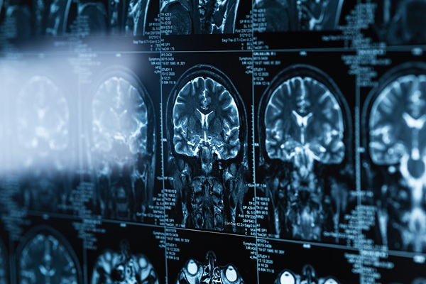
Image: Color-enhanced coronal MRI image of the brain, showing the regions of the brain most severely affected in Alzheimer’s disease (Photo courtesy of Living Art Enterprises).
In AD, nerve cell death and tissue loss cause all areas of the brain, particularly the hippocampus region, to shrink. Utilizing MRI with high spatial resolution allows radiologists to visualize slight anatomic changes in the brain that signal atrophy, or shrinkage. But the conventional practice for measuring brain tissue volume with MRI, called segmentation, is a complicated, lengthy process, according to the researchers, who published their study in the July 2008, issue of the journal Radiology.
"Visually evaluating the atrophy of the hippocampus is not only difficult and prone to subjectivity, it is time-consuming,” explained the study's lead author, Olivier Colliot, Ph.D, from the Cognitive Neuroscience and Brain Imaging Laboratory (Paris, France). "As a result, it hasn't become part of clinical routine.”
In the study, the researchers used an automated segmentation process with computer software developed in their laboratory by Marie Chupin, Ph.D., to measure the volume of the hippocampus in 25 patients with Alzheimer's disease, 24 patients with mild cognitive impairment, and 25 healthy older adults. The MRI volume measurements were then compared with those reported in studies of similar patient groups using the visual, or manual, segmentation method.
The researchers found a significant reduction in hippocampal volume in both the AD and cognitively impaired patients when compared to the healthy adults. Alzheimer's patients and those with mild cognitive impairment had a median volume loss in the hippocampus of 32% and 19%, respectively. Studies utilizing manual segmentation methods have reported similar results. "The performance of automated segmentation is not only similar to that of the manual method, it is much faster,” Dr. Colliot said. "It can be performed within a few minutes versus an hour.”
One of the goals of modern neuroimaging is to help in the early and accurate diagnosis of Alzheimer's disease, which can be challenging. When the disease is diagnosed early, drug treatment can help improve or stabilize patient symptoms. "Combined with other clinical and neuropsychological evaluations, automated segmentation of the hippocampus on MR images can contribute to a more accurate diagnosis of Alzheimer's disease,” Dr. Colliot said.
Related Links:
Cognitive Neuroscience and Brain Imaging Laboratory

Post-Processing Imaging System
DynaCAD Prostate

Breast Localization System
MAMMOREP LOOP

MRI System
nanoScan MRI 3T/7T

Half Apron
Demi