MRI-Based AI Tool Supports Differentiation of Parkinsonian Syndromes
Posted on 10 Apr 2026
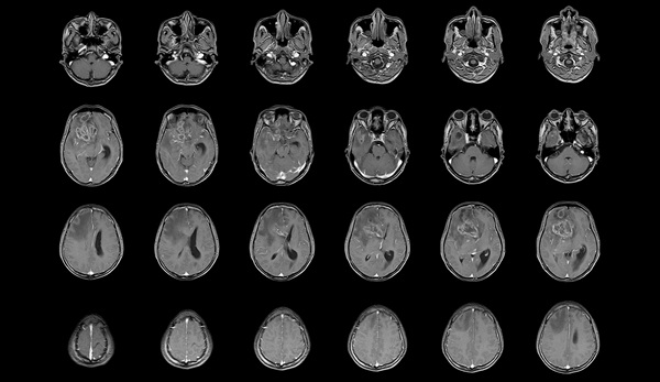
Clinicians often struggle to differentiate Parkinsonian syndromes at initial presentation, when symptom overlap can obscure disease trajectory and delay targeted care. Imaging markers derived from diffusion magnetic resonance imaging may provide objective evidence to augment clinical evaluation. A new system has launched with U.S. regulatory classification that offers AI-based support for distinguishing atypical parkinsonian disorders from Parkinson’s disease using standard MRI.
Neuropacs Corp. has announced that the U.S. Food and Drug Administration (FDA) has granted De Novo classification to neuropacs, a diffusion MRI–based software application intended to assist in the evaluation of Parkinsonian syndromes. The designation establishes the first FDA category described as a “Parkinsonian syndrome diagnostic aid.” The software provides supplemental information to neuroradiologists and neurologists as part of a comprehensive clinical assessment rather than functioning as a standalone diagnostic.
")
The system analyzes diffusion MRI data to generate a classification report based on degenerative brain patterns of multiple system atrophy, Parkinsonian variant (MSAp), and progressive supranuclear palsy (PSP), supporting differentiation from Parkinson’s disease. The analysis uses a rigorously validated free‑water imaging model in combination with a machine learning algorithm. It operates with standard diffusion MRI sequences commonly used on 3 Tesla (3T) clinical MRI systems from Siemens, GE Healthcare, and Philips, and offers cloud‑based analysis integrated into existing MRI workflows.
The technology is built on more than 15 years of research encompassing over 1,000 imaging datasets. In a prospective multicenter study published in JAMA Neurology and supported by the National Institutes of Health, the approach demonstrated performance in differentiating MSAp and PSP from Parkinson’s disease under defined study conditions. The study drew on a network of 21 movement disorder centers within the Parkinson Study Group, representing a large evaluation of diffusion MRI–based biomarkers in these syndromes.
Related Links
Neuropacs Corp.