New Ultrasound Technique Enables Safer Vein Access in Critically Ill Patient
Posted on 18 Sep 2025
Cannulation, the insertion of catheters into major veins near the collarbone, is a common but risky procedure that can cause complications such as pneumothorax or arterial injury. While ultrasound scanning techniques are used to improve accuracy, conventional methods often fail to provide a clear anatomical view, increasing the risk of misplacement. Now, a new ultrasound-guided approach for infraclavicular vein cannulation has been shown to improve both safety and success in critically ill patients.
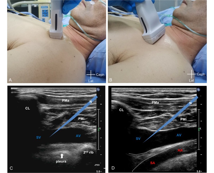
The new method, developed by researchers at Shanghai Jiao Tong University School of Medicine (Shanghai, China), is called Seeing Artery and Vein Simultaneously in the long axis (SAVES). By positioning the ultrasound probe infraclavicularly, SAVES provides a longitudinal view of both the axillary/subclavian vein and artery, while keeping the pleura out of sight. This technique leverages the anatomical space created by the anterior scalene muscle, reducing the risk of damaging critical structures during insertion.

A retrospective study analyzed consecutive critically ill adults undergoing infraclavicular axillary/subclavian vein cannulation with SAVES in a 12-bed ICU between August 2021 and December 2024. Results from 142 punctures in 111 patients demonstrated a 100% overall success rate and a 75.4% first-pass success rate. Complications were minimal, with hematoma formation at 2.1%, catheter malposition at 5.6%, and no cases of pneumothorax, hemothorax, arterial puncture, or cardiac tamponade.
The findings, published in the Journal of Intensive Medicine, demonstrated that SAVES offers clear advantages over classical ultrasound methods, which typically visualize only one vessel and include the pleura in view. By simultaneously displaying the vein and artery while keeping the pleura excluded, the technique minimizes risks of iatrogenic injury and misplacement. While current results are highly promising, researchers emphasize the need for larger prospective studies to validate the findings and support broader clinical adoption.
“SAVES technique may be a safe and effective approach for ultrasound-guided infraclavicular axillary/subclavian vein cannulation,” said Dr. Lei Li, lead author of the study.


