MRI-Based Imaging Technique Enables Rapid Assessment of Ovarian Cancer Subtypes and Treatment Response
Posted on 10 Dec 2024
Ovarian cancer patients often have multiple tumors spread across their abdomen, making it difficult to biopsy all of them, especially since they may belong to different subtypes that respond differently to treatments. Current testing methods usually result in patients waiting weeks or even months to learn whether their cancer is responding to treatment. Now, a new MRI-based imaging technique can predict how ovarian cancer tumors will respond to treatment and provide rapid feedback on the therapy's effectiveness using patient-derived cell models.
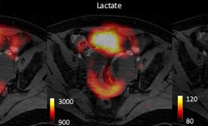
This innovative technique, developed by scientists at the University of Cambridge (Cambridge, UK), is known as hyperpolarized carbon-13 imaging. It amplifies the MRI signal by more than 10,000 times, allowing for more detailed observation. The technique works by using an injectable solution that contains a labeled form of pyruvate, a naturally occurring molecule. Once injected, the pyruvate enters the body’s cells, and the MRI scan detects how quickly it is metabolized into lactate. The rate of this metabolic process helps reveal the tumor’s subtype and its sensitivity to treatment. The researchers found that hyperpolarized carbon-13 imaging could distinguish between two ovarian cancer subtypes, providing insight into their treatment responses. They used this method to examine patient-derived cell models that closely replicate the behavior of high-grade serous ovarian cancer, the most common and lethal type of the disease.

This imaging technique can clearly identify whether a tumor is sensitive or resistant to Carboplatin, a common first-line chemotherapy drug for ovarian cancer. This capability allows oncologists to predict how well a patient will respond to treatment and assess the treatment’s effectiveness within the first 48 hours. The fast feedback from this technique enables oncologists to tailor and adjust treatment plans for each patient much sooner. In their study, published in the journal Oncogene, the scientists compared hyperpolarized carbon-13 imaging with Positron Emission Tomography (PET), which is widely used in clinical practice. They found that PET scans failed to detect the metabolic differences between tumor subtypes, meaning it couldn’t predict the type of tumor present. This study further supports the potential of hyperpolarized carbon-13 imaging for broader clinical use, and the next step will involve testing the technique in ovarian cancer patients, which the researchers expect to begin in the next few years.
“This technique tells us how aggressive an ovarian cancer tumor is, and could allow doctors to assess multiple tumors in a patient to give a more holistic assessment of disease prognosis so the most appropriate treatment can be selected,” said Professor Kevin Brindle in the University of Cambridge’s Department of Biochemistry, senior author of the report. “We can image a tumor pre-treatment to predict how likely it is to respond, and then we can image again immediately after treatment to confirm whether it has indeed responded. This will help doctors to select the most appropriate treatment for each patient and adjust this as necessary.”


