Collaboration to Make Diagnostic Medical Imaging Less Hazardous Using Hyperpolarization Agents
By MedImaging International staff writers
Posted on 23 Oct 2014
A collaborative effort by scientists has led to the development of an innovative strategy that can considerably improve the capabilities of medical imaging with safer procedures for the patient.Posted on 23 Oct 2014
Medical imaging is a significant part of healthcare today, with imaging techniques such as magnetic resonance imaging (MRI), computed tomography (CT) scanning, and nuclear magnetic resonance (NMR) increasing greatly over the last 20 years. However, continuing problems of image resolution and quality still hinder these techniques because of the nature of living tissue. A solution is hyperpolarization, which involves injecting the patient with substances that can enhance imaging quality by following the distribution and fate of specific molecules in the body but that can be harmful or potentially toxic to the patient.

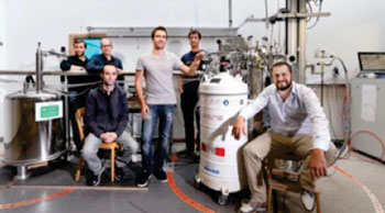
Image: A collaborative effort between EPFL, CNRS, ENS Lyon, CPE Lyon, and ETH Zürich has led to the development of a novel approach that can considerably improve the capabilities of medical imaging with safer procedures for the patient (Photo courtesy of EPFL - Ecole Polytechnique Fédérale de Lausanne).
A team of scientists from Ecole Polytechnique Fédérale de Lausanne (EPFL; Switzerland), Le Centre National de la Recherche Scientifique (CNRS; Paris, France), ENS Lyon ( École Normale Supérieure de Lyon; France), and CPE Lyon (France), and Swiss Federal Institute of Technology in Zurich (ETH Zürich; Switzerland) has developed a new generation of hyperpolarization agents that can be used to dramatically enhance the signal intensity of imaged body tissues without presenting any danger to the patient. Their research was published September 29, 2014, in the journal Proceedings of the National Academy of Sciences of the United States of America (PNAS).
The team of scientists coordinated by Dr. Lyndon Emsley, a professor at EPFL and ENS Lyon, has developed a new generation of hyperpolarizing agents that are both effective and safe for the patient. The substances, called HYPSOs, were developed by the teams of Christophe Copéret at ETH Zurich and Chloé Thieuleux at CPE-Lyon. The HYPSOs come in the form of a fine, white, porous powder that contains the “tracking” molecules to be hyperpolarized. The HYPSO powder is comprised of mesoporous silica (silicon dioxide), which is the major component of sand and is commonly used in nanotechnology.
The silica powder used for the HYPSOs consists of particles, containing pore channels. It has been designed in such a way that the surface of each pore channel can be evenly covered with molecules known as “organic radicals.” The radicals are homogeneously distributed, and are able to induce polarization around them. The pore channels are then filled with a solution of the “tracking” molecules to be hyperpolarized, which act as markers for the imaging—e.g., pyruvate, which is important in the production of energy in cells.
Using novel instruments and methods developed by Dr. Sami Jannin at EPFL, the HYPSO sample is hyperpolarized with microwaves in a magnetic field at a very low temperature. The magnetic moments of the atoms are forced to align through a process called “dynamic nuclear polarization,” which transfers the spin energy of the free radicals’ electrons to the markers’ nuclei. The electronic spin magnetism of the hyperpolarizing agent acts on the marker molecule, aligning, or “polarizing,” the nuclei of its atoms.
Hot water is then used to melt and flush the substrate out of the powder. Because of the equipment and conditions needed, the process generally takes place in a room adjacent to the imaging facility. The substrate is then injected through a long tube into the patient inside the medical imaging device. The entire process only lasts about 10 seconds.
Two scans are performed, one with and one without the hyperpolarized agent. When the two images are compared, it is possible to observe the distribution of the hyperpolarized marker in the patient’s body, which, depending on the medical setting, can be indicative of disease. For example, accumulation of pyruvate in the prostate could be an early indication of prostate cancer.
The researchers have evaluated the effectiveness of the HYPSOs technology on several imaging markers, including pyruvate, acetate, fumarate, pure water, and a simple peptide. Because the HYPSOs is physically retained during dissolution, the technique generates pure solutions of hyperpolarized markers, free of any contaminant. The protocol is therefore simpler and potentially safer for the patient, while its dramatic efficiency on signal quality forecasts the use of this new generation of hyperpolarized agents with a broad range of molecules. As Dr. Jannin pointed out, “We have now received queries of scientists from abroad who are eager to boost their research with this new technology. Amongst other plans, we are very excited about testing these materials in vivo.”
Related Links:
Ecole Polytechnique Fédérale de Lausanne
Le Centre National de la Recherche Scientifique
ENS Lyon


.jpg)