Special Glasses Help Surgeons Visualize Cancer
By MedImaging International staff writers
Posted on 27 Feb 2014
High-tech glasses may help surgeons distinguish cancer cells from healthy cells by making them glow blue when viewed through the eyewear.Posted on 27 Feb 2014
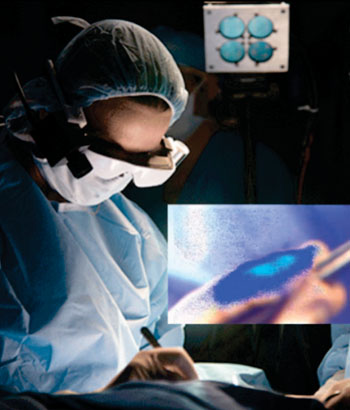
Developed by researchers at Washington University (St. Louis, MO, USA), the technology, called optical projection of acquired luminescence (OPAL), incorporates custom video technology, a head-mounted display, and a targeted molecular contrast agent that attaches to cancer cells, making them glow blue when irradiated with a special light. The resulting fluorescence intensity maps are projected onto the imaged surface and viewed through the glasses, rather than via wall-mounted display monitors.

Image: Breast surgeon Julie Margenthaler, MD, visualizing cancer cells (Photo courtesy of WUSTL - Washington University in St. Louis).
To demonstrate the proof-of-principle for OPAL applications in oncologic surgery, lymphatic transport of indocyanine green was visualized in live mice for real-time identification of sentinel lymph nodes. Subsequently, peritoneal tumors in a murine model of breast cancer metastasis were identified using OPAL following systemic administration of a tumor-selective fluorescent molecular probe. The researchers noted that tumors as small as one mm in diameter could be detected. The study was published in the December 2013 issue of the Journal of Biomedical Optics.
“These initial results clearly show that OPAL can enhance adoption and ease-of-use of fluorescence imaging in oncologic procedures relative to existing state-of-the-art intraoperative imaging systems,” said lead author professor of radiology and biomedical engineering Samuel Achilefu, PhD. “This technology has great potential for patients and health-care professionals. Our goal is to make sure no cancer is left behind.”
“We’re in the early stages of this technology, and more development and testing will be done, but we’re certainly encouraged by the potential benefits to patients,” said associate professor of surgery Julie Margenthaler, MD, who performed the first in-human operation using the system at the WUSTL Barnes-Jewish Hospital (St. Louis, MO, USA) on February 10, 2014. “Imagine what it would mean if these glasses eliminated the need for follow-up surgery and the associated pain, inconvenience, and anxiety.”
Related Links:
Washington University
Barnes-Jewish Hospital


.jpg)