Contrast-Free Dynamic Diffuse Optical Tomography Imaging for Peripheral Artery Disease Shows Potential
By MedImaging International staff writers
Posted on 18 Sep 2012
Peripheral artery disease (PAD), a narrowing of arteries in the arms and legs, is a frequent result of out of control diabetes, sometimes even leading to amputations. Symptoms such as foot pain and numbness are frequently the first signs that patients have PAD. An earlier diagnosis would be extremely helpful for thousands, long before effective intervention becomes ineffective.Posted on 18 Sep 2012
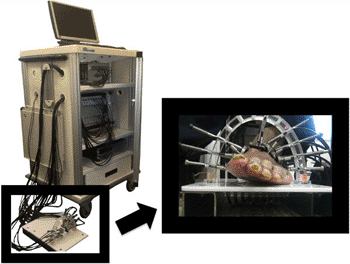
A new technique called dynamic diffuse optical tomography imaging (DDOT) used by researchers from Columbia University (New York, NY, USA) may provide to new diagnostic approach to identify PAD. DDOT uses near-infrared light to visualize hemoglobin within tissue, providing an indication of how well blood is moving below the probe. The technique does not require the injection of hazardous contrast agents nor the use of X-ray radiation, potentially helping make it suitable for a screening test.

Image: A new technique called dynamic diffuse optical tomography imaging (DDOT) is intended to provide earlier diagnosis in patients with peripheral artery disease (Photo courtesy of Columbia University).
Michael Khalil, a PhD candidate working with Andreas Hielscher, PhD, professor of biomedical and electrical engineering and radiology, and director of the biophotonics and optical radiology laboratory at Columbia. “One key reason why DDOT shows so much promise as a diagnostic and monitoring tool is that, unlike other methods, it can provide maps of oxy, deoxy, and total hemoglobin concentration throughout the foot and identify problematic regions that require intervention.”
Contrast agents pose the risk of renal failure in some cases, therefore, the ability to monitor PAD without using a contrast agent is a great advantage.
“Using instrumentation for fast image acquisition lets us observe blood volume over time in response to stimulus such as a pressure cuff occlusion or blockage,” said Prof. Hielscher. “In the case of tissue, light is absorbed by hemoglobin. Since hemoglobin is the main protein in blood, we can image blood concentrations within the foot without using a contrast agent.”
Related Links:
Columbia University


