Special Nanoparticles That Could Serve As Contrast Agents to Improve Modern Imaging Techniques
Posted on 01 Nov 2023
Researchers have developed special nanoparticles that have the potential to enhance existing imaging technologies. These special nanoparticles respond to heat, altering their properties. When combined with an integrated dye, these particles could be a game-changer for photoacoustic imaging, producing detailed, three-dimensional internal images of the human body.
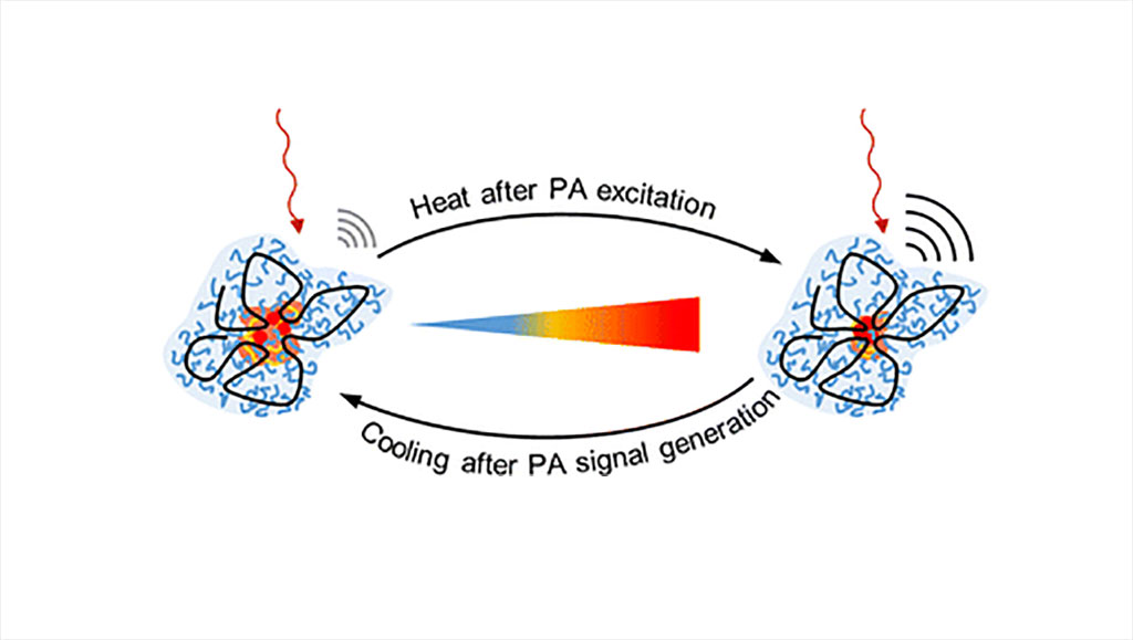
Researchers at Martin Luther University Halle-Wittenberg (MLU, Halle, Germany) have engineered single-chain nanoparticles (SCNPs), incredibly small structures that are just three to five nanometres in size and comprised of a single molecular chain. These tiny capsules allow dyes to be incorporated within them. What sets these SCNPs apart is their unique thermoresponsive nature: their structure changes when they come in contact with heat. Depending on the temperature, their structure can alternate between a compact and an open configuration. This change also affects the behavior of the substances enclosed within the particles.

For the study, the team integrated special dyes into these SCNPs for use in photoacoustic imaging. In this technique, laser beams are aimed at the tissue for examination. The laser light then gets converted into ultrasound waves, causing the tissue to warm up and the nanoparticles to alter their properties. Capturing these ultrasound waves from outside the body allows for the creation of three-dimensional images, predominantly revealing blood vessel structures. According to the scientists, the nanoparticles contribute to a strong optical contrast that could be particularly useful in tumor analysis. The researchers also explored the effectiveness of these particles in cell cultures to gain insights into how they might function within the human body. Such understanding is vital if these nanoparticles are to find their way into biomedical uses. The newly-developed particles excelled in all the tests they were subjected to.
"Our work is an important step in the development of thermoresponsive SCNPs, which could improve the accuracy and precision of diagnostic imaging," said chemist Professor Wolfgang Binder from MLU, who led the study.
Related Links:
MLU


