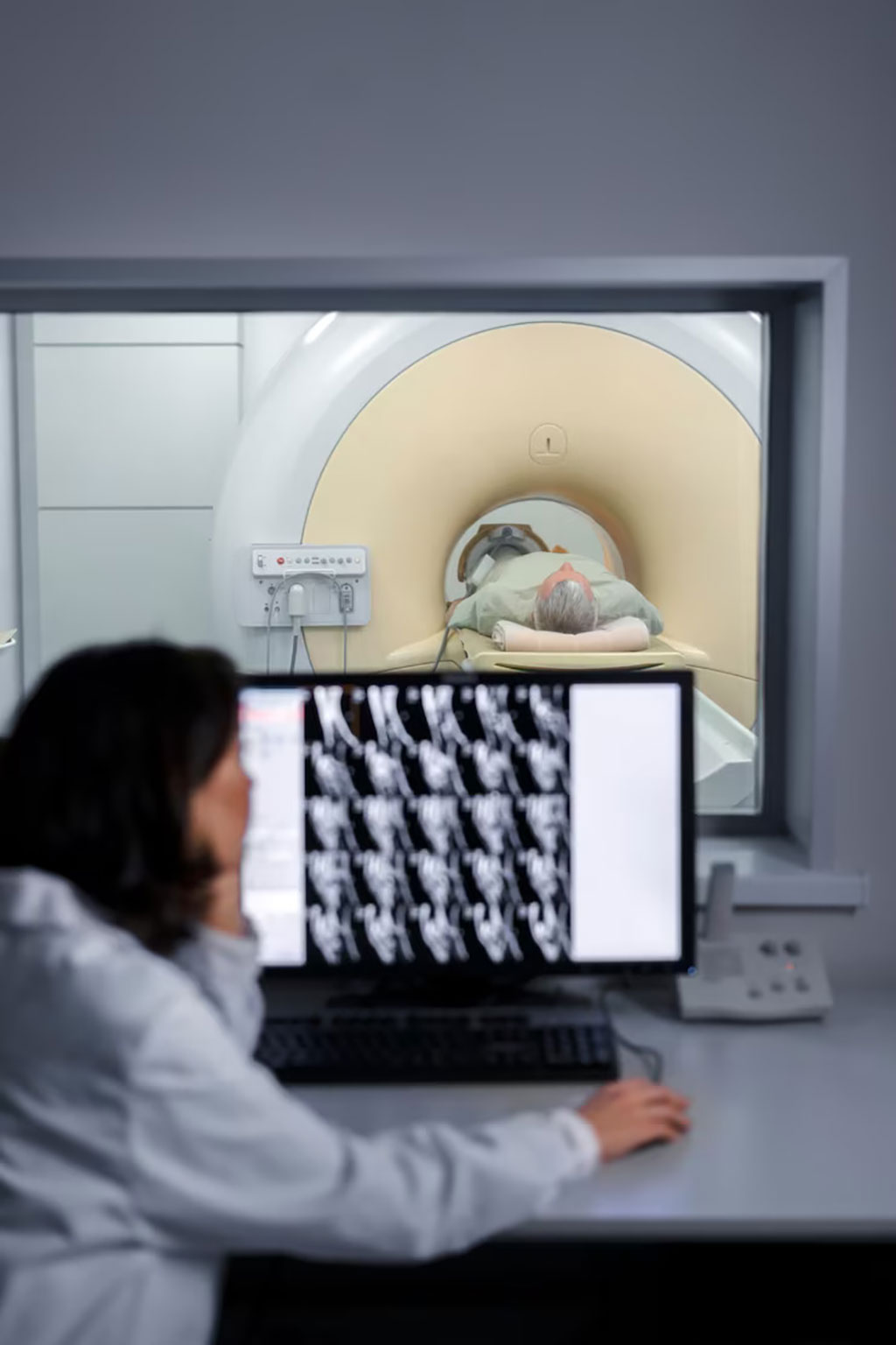
New Use for Copper in MRI Contrast Agent Design Enables Clearer Images and Improved Diagnosis
Posted on 12 Jul 2023
Magnetic resonance imaging (MRI) scanners illuminate portions of the body using a strong magnetic field, leading to hydrogen nuclei of water in tissues being polarized in the direction of the magnetic field. The magnitude of the spin polarization is measured to generate MR images but decays according to a specific time constant known as the T1 relaxation time. Water protons in different tissues exhibit varied T1 values, serving as a primary source of contrast in MR images. The T1 value of nearby water protons can be both reduced and, occasionally, increased through the use of a contrast agent, enhancing the image contrast and thereby boosting the clarity of internal bodily structures. The most common compounds used for this purpose are gadolinium-based contrast agents (GBCAs). While Gadolinium (in the form of Gd3+) is frequently employed as a contrast agent, environmental and patient safety concerns have led to the ongoing search for new contrast agents.
In a groundbreaking research collaboration, scientists at the University of Birmingham (Birmingham, UK) have uncovered a novel application of copper in designing MRI contrast agents. This finding holds promise for generating superior, safer images that facilitate easier and safer patient diagnosis. The researchers identified a novel copper protein binding site that holds significant potential for use in MRI contrast agents to enhance the visibility of internal body structures. This discovery defies the traditional belief that copper is ill-suited for MRI contrast agents and may contribute to the creation of new imaging agents posing fewer risks and side effects than those currently in use.

The research team succeeded in creating a highly elusive abiological copper site bound to oxygen donor atoms within a protein scaffold. They found that this new structure exhibited high levels of relaxivity - the capacity of a contrast agent to affect the proton relaxation times, leading to clearer and more detailed MRI images. The researchers suggest that copper-based imaging agents may also be used in Positron emission tomography (PET) scans, which generate intricate 3D internal body images. Their study highlights how the creation of a copper site within a protein scaffold using an artificial coiled coil resulted in functionality and performance not typically linked to copper.
“We prepared a new-to-biology copper–binding site which shows real potential for use in contrast agents and challenges existing dogma that copper is unsuitable for use in MRI,” said co-author Dr Anna Peacock, Reader in Bioinorganic Chemistry at the University of Birmingham. “Despite copper largely being disregarded for use in MRI contrast agents, our binding site was shown to display extremely promising contrast agent capabilities, with relaxivities equal and superior to the Gd(III) agents used routinely in clinical MRI. Our discovery showcases a powerful approach for accessing new tools or agents for imaging applications.”
Related Links:
University of Birmingham


