Molecular Imaging Technique Improves Endometriosis Diagnosis
Posted on 08 Jun 2022
Endometriosis is a common inflammatory condition affecting women, associated with painful periods, chronic pelvic pain, and infertility. It is believed to be due to tissue that resembles the lining of the womb sticking to areas outside the uterus, usually in the pelvis. However, diagnosing endometriosis often results in multiple visits to physicians, multiple scans and often surgery to confirm the diagnosis. As a result, there is a delay to get a diagnosis of endometriosis, mainly due to lack of non-invasive tests capable of detecting all endometriosis subtypes (ovarian, superficial, and deep disease). Now, a new research study is investigating whether a 20-minute imaging scan can detect the most common types of endometriosis, which currently require surgery to diagnose.
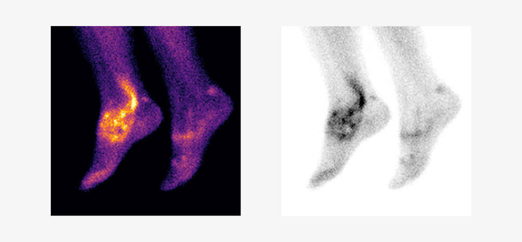
Endometriosis experts at the University of Oxford (Oxford, UK) have joined hands with Serac Healthcare Ltd. (London, UK) to investigate whether an experimental imaging marker (⁹⁹ᵐTc maraciclatide) that binds to areas of inflammation can be used in endometriosis to visualize the disease on a scan. The imaging marker has been used for detecting inflammation in conditions such as rheumatoid arthritis in other research studies across the UK, EU and US. Maraciclatide is for investigational use only and is not approved by the FDA or UK and European regulatory authorities.

The potential strengths of the scan lie in the way the imaging marker binds to areas of inflammation, which may allow doctors to distinguish between new and old lesions and detect endometriosis in areas not easily seen during surgery such as the lung. The development of a 20-minute imaging test would reduce the need for repeated visits to doctors, for repeated investigations, and for invasive surgery to obtain a diagnosis. This would ultimately reduce the time taken to confirm or exclude endometriosis.
Initially, the study will involve women who are scheduled to have planned surgery for suspected endometriosis. Two to seven days before their operation, the participants will be invited for an imaging scan which will compare the suspected locations of disease detected on the scan and in surgery to confirm whether this imaging test could be an effective non-invasive method of detecting all endometriosis subtypes.
“There is an urgent unmet clinical need for a non-invasive marker to identify or rule out endometriosis as it is such a very common disease affecting more than 190 million women worldwide,” said Professor Christian Becker, Co-Director of the Endometriosis CaRe Centre in Oxford, who will lead the study.
“We are excited about the potential of ⁹⁹ᵐTc maraciclatide to diagnose endometriosis non-invasively and delighted to be working with the internationally renowned team at Oxford on this important first study,” said David Hail, CEO of Serac Healthcare.
Related Links:
University of Oxford
Serac Healthcare Ltd.


