Medical Holography System Creates Spatially Accurate Images
By MedImaging International staff writers
Posted on 16 Aug 2021
A new three-dimensional (3D) visualization system allows direct interaction with floating holograms of a patient's true anatomy, based on standard CT scans and 3D ultrasonography. Posted on 16 Aug 2021
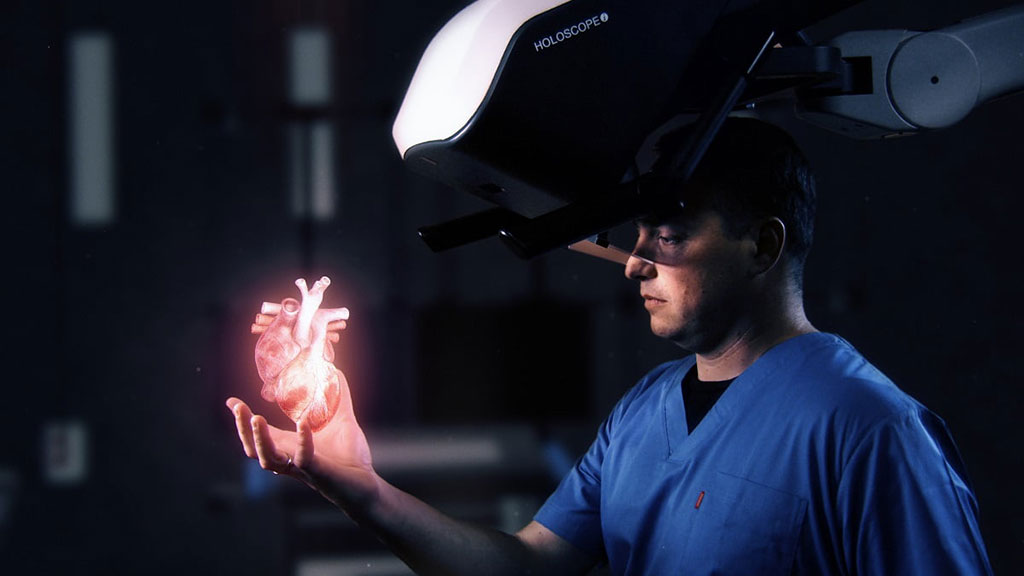
The RealView Imaging (Yokneam, Israel) HOLOSCOPE-i system uses an ‘over-the-head’ projection system suspended above the physician, without the need for any head-mounted device, special eyewear, or interaction tools. Using proprietary Digital Light Shaping (DLS) technology, true interference-based volumetric holography (a Nobel Prize winning technology) is made available, creating holograms with high spatial resolution in all three dimensions. The unique technological advantage allows natural 3D/4D visualization and prolonged use of the system without provoking fatigue, nausea, or headache.

Image: The HOLOSCOPE-I over-the-head holographic system (Photo courtesy of RealView Imaging)
The digital data required to create the hologram is imported from a PACS, USB, or directly from a 3D ultrasound scanner to the RealView algorithms on the system computer. The resulting output is a digital interference pattern that is projected onto a spatial light modulator (SLM). The hologram is then created by twin optical channels consists of an SLM, a red-green-blue (RGB) coherent light source, and a set of lenses and mirrors to direct the light propagation, ending with the see-through eyepieces that enable the viewer to see the 3D hologram at arms-reach.
The software-controlled system is comprised of the twin SLM optical projection units; a computer and electronics unit that support human machine interactions (HMI) and a graphical user interface (GUI) display; a cart and boom mechanical fixture connecting the SLM optical unit and the system computer; and a 3D controller for interfacing with the hologram. The resulting hologram, floating in air, provides a 3D image of the data and enables visualization of the image and interaction with it, such as touching, marking, locating, slicing, or defining a pathway for the intervention.
“Having a real hologram of the heart in my hand, based on pre-operative CT and intra-procedure ultrasound, allows me to focus-in and fully understand the complexities of the patient's 3D anatomy,” said Elchanan Bruckheimer, MD, medical director of RealView Imaging. “I can intuitively comprehend the dynamic spatial anatomical relationships of the cardiac valve leaflets, for example. This technology provides me with more confidence, potentially resulting in shorter procedures and better outcomes.”
The company's next product, the HOLOSCOPE-x, will project 3D holographic images inside the closed patient's body (in situ), making the patient’s anatomy literally transparent, enabling spatially-precise minimally-invasive procedures.
Related Links:
RealView Imaging


