Mobile Devices Prove Reliable for Acute Stroke Diagnosis
By MedImaging International staff writers
Posted on 25 Feb 2020
A new study shows that interpretation of a computerized tomography (CT) scan on a smartphone or laptop is reliable prior to IV thrombolysis administration in acute stroke patients. Posted on 25 Feb 2020
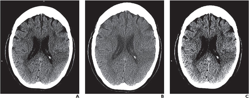
Researchers at the University of Los Andes (UniAndes; Bogotá, Colombia), Hospital Fundación Santa Fe de Bogotá (Colombia), and the Baptist Neurological Institute (Jacksonville, FL, USA) conducted a retrospective study of 2,256 CT interpretations to evaluate the reliability of head CT images displayed on smartphone or laptop reading systems in 880 patients with symptoms of acute stroke, compared with those made after interpretation of images displayed on a medical workstation monitor.

Image: A CT image as seen on a Barco E-2620 monitor (A), Samsung Galaxy S8 Plus smartphone (B), and a Lenovo ThinkPad T460s laptop (C) (Photo courtesy of AJR)
The CT interpretations on all three systems, conducted by four neuroradiologists, were then analyzed to calculate intra-observer and inter-observer agreements using the intraclass correlation coefficient (ICC) and interpretation variables that included hemorrhagic lesions; infarct size assessment; stroke dating (acute, subacute, and chronic); intraaxial neoplasm; and hyperdense arteries. In addition, accuracy equivalence tests were performed on recommendations for IV thrombolysis with recombinant tissue plasminogen activator (tPA).
The results showed that good or very good intra-observer agreements were shown for all the variables, and specifically for those variables required to establish contraindications for IV thrombolysis, where the agreements were ranked as very good. The tPA IV thrombolysis recommendation showed very good inter-observer agreements, with AUC values and sensitivities equivalent among all the reading systems at a 5% equivalent threshold. The study was published on January 28, 2020, in the American Journal of Roentgenology (AJR).
“Our study found that mobile devices are reliable and accurate to help stroke teams to decide whether to administer IV thrombolysis in patients with acute stroke,” concluded lead author Antonio Salazar, PhD, of the UniAndes Laboratory of Telemedicine and Electrophysiology, and colleagues. “These results constitute a strong foundation for the development of mobile-based telestroke services, because they increase neuroradiologist availability and the possibility of using reperfusion therapies in resource-limited countries.”
Mobile phone screen resolution has been significantly increased over the years, from the incipient QCIF (176 × 144 pixel) standard to the current Quad HD (2560 × 1440 pixel) or even Ultra HD (3840 × 2160 pixel) resolutions. At the same time, screen size has also been enlarged, from smaller than one inch to seven inches, or even larger. But the most important features for accurate medical grade images is the number of colors on grey scale images, contrast modulation, viewing angle, and calibration.
Related Links:
University of Los Andes
Hospital Fundación Santa Fe de Bogotá
Baptist Neurological Institute


