Fluorescent Markers Illuminate Cancer Intraoperatively
By MedImaging International staff writers
Posted on 19 Feb 2020
A new study shows that intraoperative fluorescent markers can be used to illuminate and target non-small-cell lung cancer (NSCLC), improving patient outcomes. Posted on 19 Feb 2020
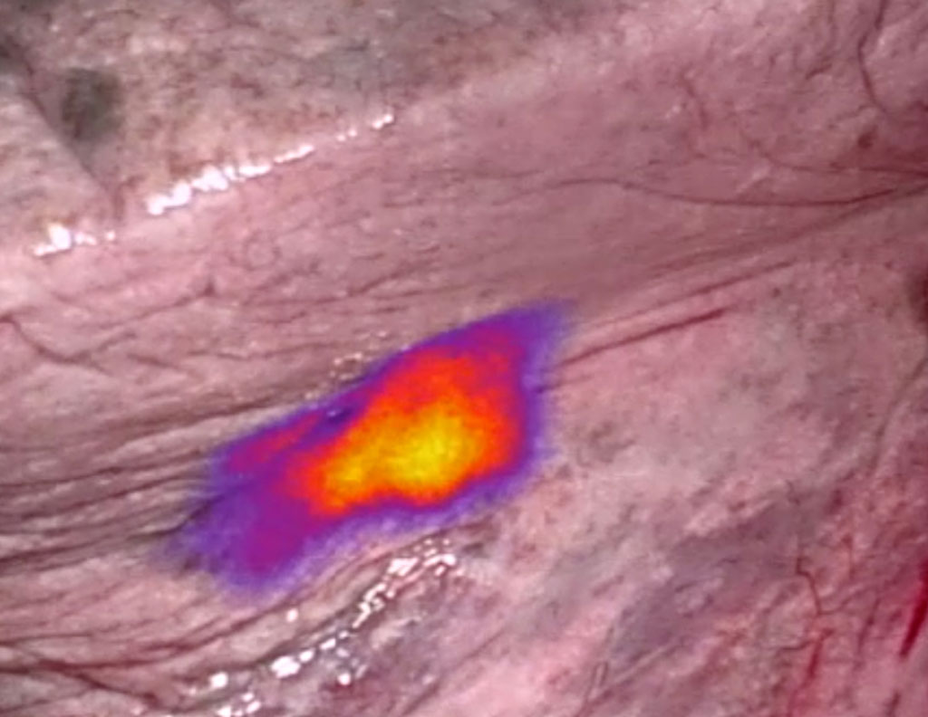
Under development at On Target Laboratories ((Lafayette, IN, USA), based on technology developed by Purdue University (Lafayette, IN, USA), OTL38 is an analog of folate (vitamin B9) conjugated to a near-infrared (NIR) fluorescent dye. A small single dose of the compound is administered via intravenous (IV) infusion prior surgery. A dedicated near-infrared (NIR) imaging system is then used to identify the fluorescent signal generated after the agent binds to folate receptor-alpha (FRα). Since NIR light at 796 nm penetrates several centimeters into tissue, OTL38 can visualize tumors under the surface.

Image: Fluorescent markers help identify and remove cancer cells during surgery (Photo courtesy of On Target)
In a multi-institutional Phase 2 clinical trial involving 92 patients with NSCLC conducted over 18 months, OTL38 improved outcomes for 26% of patients undergoing pulmonary resection for NSCLC, and helped uncover otherwise unlocalizable lesions in 11 patients (12%). In addition, 10 supplementary cancers were found in seven patients, (8%), and inadequate surgical margins were identified in eight patients (9%). The study was presented at the 56th annual meeting of the Society of Thoracic Surgeons (STS), held during January 2020 in New Orleans (LA, USA).
“OTL38 is the first technique that is specific to imaging adenocarcinomas of the lung, which is one of the most common types of invasive lung cancer,” said cardiothoracic surgeon Inderpal Sarkaria, MD, of the University of Pittsburgh Medical Center (PA, USA). “NIR imaging with OTL38 may be a powerful tool to help surgeons significantly improve the quality of lung cancer surgery by more clearly identifying tumors and allowing the surgeon to better see and completely remove them, one of the most vital components in the overall care of patients with this disease.”
Folate can be used like a Trojan horse to sneak an imaging agent or drug into a cancer cell. About 80% of endometrial, lung, kidney, and ovarian cancer exhibit high rates of FR-α receptor expression, and 50% of breast and colon cancers also express the receptor.
Related Links:
On Target Laboratories
Purdue University


