Skull Clamp Supports Advanced Head and Neck Surgical Imaging
By MedImaging International staff writers
Posted on 09 Jan 2020
A novel intraoperative skull clamp optimizes head and neck visualization for image-guided surgery, deep brain stimulation (DBS) and other interventions in the operating room (OR) environment.Posted on 09 Jan 2020
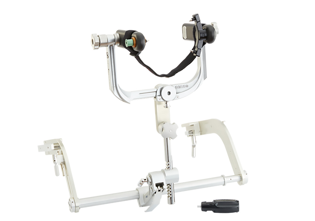
The NeuroLogica (Danvers, MA, USA) DORO QR3 XTom intraoperative skull clamp is designed to support the OmniTom Mobile 16-Slice CT scanner. Features include exceptional stability through an easy-to-use parkbench mount and additional stabilization to the table’s siderail, ultra-precise clamp adjustment, radiolucent extension arms for fine-tuning patient placement, and an ultra-wide quick rail that accommodates brain tissue retractor systems. The unit provides two interfaces for image-guided brain navigation systems, and dismantles easily to simplify disinfection.

Image: The DORO QR3 XTom intraoperative skull clamp (Photo courtesy of NeuroLogica)
The OmniTom Mobile 16-Slice CT scanner features a gantry opening of 40 cm that allows improved coverage of the adult head and neck area and full body pediatric scanning. Advanced reconstruction is available for 3D and multiplanar imaging, mean slab, maximum/minimum intensity projection, and oblique datasets. Metal artifact and other corrections can also be added to the primary reconstruction, including noise reduction, Iodine delivery rate (IDR), direct digital radiography (DDR), windmill artifact reduction, automatic bolus tracking and contrast injector triggering to maximize workflow efficiency.
The 16-slice (0.625 mm per slice) advanced data acquisition system offers effective dose optimization and highly advanced N-DAS noise detectors which were combined with a 24-bit processing chain that never compresses the data, leading to clearer images with ultimately low artificial noise. In addition, automatic exposure control (AEC) provides mA modulation during helical and axial scanning in order to regulate dose and image quality. It also features an array of mobility features, including an internal drive system and smart-sensing collision avoidance software.
“The OmniTom brings state-of-the-art head and neck imaging to any setting to collect patient data in real time. In the OR, with the new DORO intraoperative head clamp, the high resolution 16-slice scanner can be used for a wide range of today’s most sophisticated brain interventions,” said David Webster, chief operating officer of NeuroLogica. “By supporting precise patient positioning, the clamp enables physicians to take full advantage of the unit’s 40 cm bore with a large field of view. The clamp also provides support for today’s important surgical and imaging tools.”
Related Links:
NeuroLogica


