New MRI Technique Improves Breast Cancer Screening
By MedImaging International staff writers
Posted on 20 Mar 2019
A new magnetic resonance imaging (MRI) waistcoat packed with flexible radio frequency (RF) coils could screen women for breast cancer faster and with less disadvantages, claims a new study.Posted on 20 Mar 2019
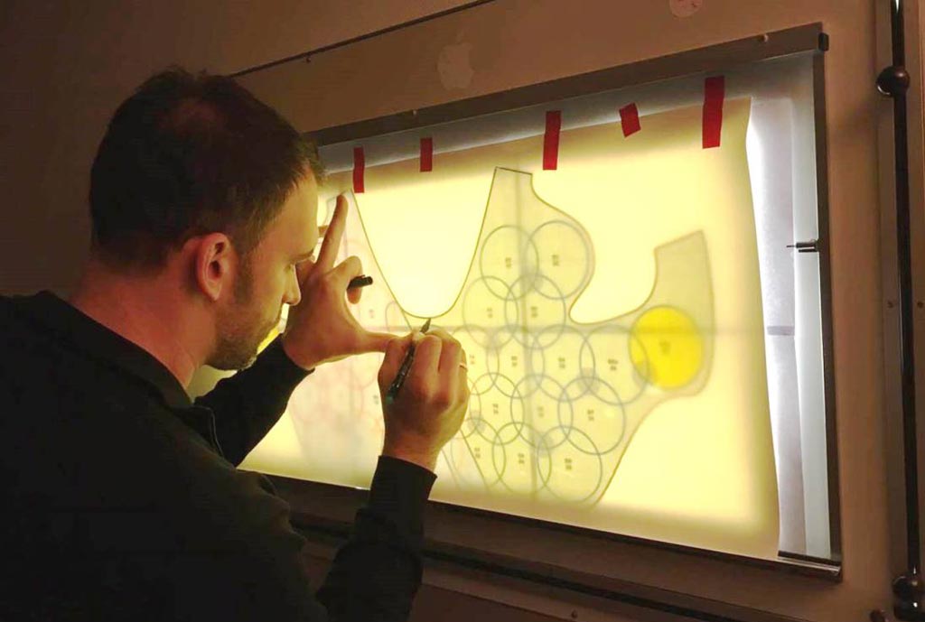
Under development at the Medical University of Vienna (MedUni; Austria), Université de Lorraine (Nancy, France), and other institutions, the prototype waistcoat device is a mechanically flexible, receive-only array that contains 32 RF coils sewn into the fabric, each consisting of an 18 centimeter long coaxial cable formed into an eight centimeter wide loop. A receiver unit at the end of the cable feeds the signal into the computer. Motion sensors built into the vest monitor the patient so that it can cancel out breathing movements that distort the image.

Image: A pattern for a MRI vest with 32 RF coils sewn in (Photo courtesy of Elmar Laistler/ MedUni).
During the examination, the woman lies face up so that the breast is flattened, exposing larger sections to the receiver, thus helping to enhance signal potency and shorten measuring time, regardless of the size of the breast. In addition, as a biopsy may often be required after the MRI examination, having the patient in prone facilitates tumor removal. In phantom and in vivo MRI experiments, an increase in signal-to-noise ratio (SNR) was demonstrated in regions close to the array and for high parallel imaging acceleration factors. A study describing the MRI waistcoat was published on November 1, 2018, in PLOS One.
“One advantage of examining the breast with MRI is the avoidance of X-rays, as used in classical mammography. The risks involved in such ionizing radiation are of concern, in particular for young women,” said senior author Elmar Laistler, PhD, of MedUni. “Further advantages of MRI are significantly higher sensitivity and better resolution of the measurement. MRI examination is well on its way to becoming the standard method, but some issues remain.”
“In MRI, the actual image is always recorded with radio frequency coils. So far, MR mammography has involved having women lie face down in the MR scanner tube,” concluded Dr. Laistler. “These coils are housed in two one-size-fits-all cups. The patient lies on her stomach so that her breasts settle into these cups. This does not work equally well for all women and all breast sizes, because the coils are more efficient and give better results depending on whether they are a good fit for the respective body shape.”
While breast MRI provides a very high sensitivity and negative predictive value, particularly in non-calcified breast lesions, problem-solving definitions are not well defined, and the empirical evidence about specific indications, such as architectural distortions, is sparse.
Related Links:
Medical University of Vienna
Université de Lorraine


