High-Resolution Microscopy Method Rapidly Maps the Brain
By MedImaging International staff writers
Posted on 30 Jan 2019
A new three-dimensional (3D) imaging technique can locate individual neurons, trace connections between them, and visualize organelles inside neurons, according to a new study.Posted on 30 Jan 2019
The technique, developed at the Massachusetts Institute of Technology (MIT, Cambridge, MA, USA), the University of California (UC, Berkeley, USA), the Howard Hughes Medical Institute (HHMI; Ashburn, VA, USA), and other institutions, combines expansion microscopy and lattice light-sheet microscopy to image the nanoscale spatial relationships between proteins across entire sections of the brain.

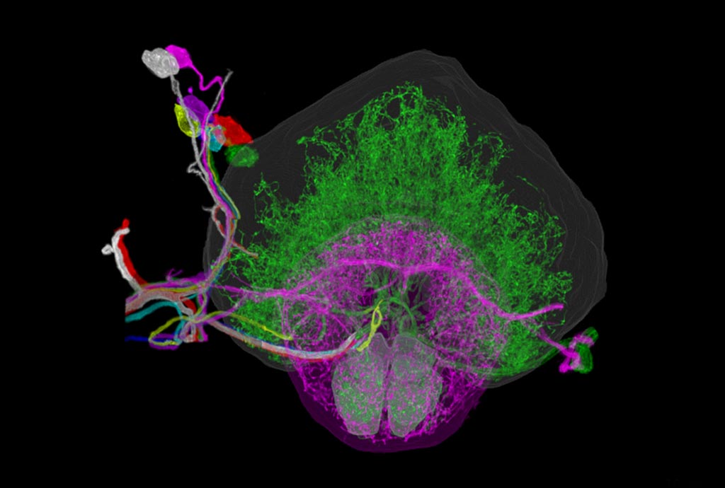
Image: The right hemisphere of a fruit fly brain (Photo courtesy of MIT).
For the study, the researchers imaged the Drosophila fly brain, including synaptic proteins at dendritic spines, myelination along axons, and presynaptic densities at dopaminergic neurons in every fly brain region. The researchers were able to compute the thickness of the myelin coating in different segments of axons, and measured the gaps between stretches of myelin. The technology can also be used to image tiny organelles inside neurons, such as mitochondria and lysosomes. The study was published on January 17, 2019, in Science.
“The marrying of the lattice light-sheet microscope with expansion microscopy is essential to achieve the sensitivity, resolution, and scalability of the imaging that we're doing. Using this technique, it is possible to analyze millions of synapses in just a few days,” said lead author Ruixuan Gao, PhD, of MIT. “We counted clusters of postsynaptic markers across the cortex, and we saw differences in synaptic density in different layers of the cortex. Using electron microscopy, this would have taken years to complete.”
“This technique allows researchers to map large-scale circuits within the brain while also offering unique insight into individual neurons' functions,” said senior author Professor Edward Boyden, PhD, of the MIT Institute for Brain Research, Media Lab, and the Koch Institute for Integrative Cancer Research. “A lot of problems in biology are multiscale. Using lattice light-sheet microscopy, along with the expansion microscopy process, we can now image at large scale without losing sight of the nanoscale configuration of biomolecules.”
Expansion microscopy starts with a preserved specimen, such as a thin slice of tissue. It is infused with an absorbent polymer that expands the sample tissue fourfold. The process also turns the sample nearly transparent, which makes it particularly well-suited for the lattice light-sheet microscope, which shines light from one side and takes a picture from the other side. According to the researchers, the technology should enable statistically rich, large-scale studies of neural development, sexual dimorphism, degree of stereotypy, and structural correlations to behavior or neural activity, all with molecular contrast.
Related Links:
Massachusetts Institute of Technology
University of California
Howard Hughes Medical Institute


