MRI Implant Monitors Biophysical Processes in Brain
By MedImaging International staff writers
Posted on 07 Nov 2018
A new study describes how a minimally invasive implantable magnetic resonance imaging (MRI) sensor can detect electrical activity or optical luminescence in the brain.Posted on 07 Nov 2018
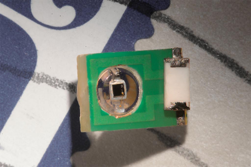
Developed at the Massachusetts Institute of Technology (MIT, Cambridge, MA, USA), the new sensor is basically a tiny antenna that detects radio waves emitted by the nuclei of hydrogen atoms in water present in brain tissues. The inductively coupled resonant sensor is initially tuned to the radio frequency emitted by the hydrogen atoms. When a local electromagnetic or photonic event occurs, the MRI signal of the sensor is modified, no longer matching the frequency of the hydrogen atoms. The coil-based transducers can then be detected via an MRI scanner, enabling the remote sensing of biological fields.

Image: A miniature sensor can measure optical and electrical signals in the brain using MRI (Photo courtesy of Felice Frankel/ MIT).
The sensor requires neither onboard power nor wired connectivity, and is sensitive enough to detect potentials on the millivolt scale, comparable to what biological tissue generates, especially in the brain. It can thus detect electrical impulses fired by single neurons (action potentials), or local field potentials produced by a group of neurons. The sensor can also detect light emitted by cells engineered to express the protein luciferase, allowing researchers to determine if the genes were successfully incorporated by measuring the light produced. The study was published on October 22, 2018, in Nature Biomedical Engineering.
“If the sensors were on the order of hundreds of microns, which is what the modeling suggests is in the future for this technology, then you could imagine taking a syringe and distributing a whole bunch of them and just leaving them there,” said senior author Professor Alan Jasanoff, PhD. “What this would do is provide many local readouts by having sensors distributed all over the tissue. It could also be used to monitor electromagnetic phenomena elsewhere in the body, including muscle contractions or cardiac activity.”
Biological electromagnetic fields arise throughout all tissue depths and types, and are associated with physiological processes and signaling in organs of the body. Currently, the most accurate way to monitor electrical activity in the brain is by inserting an electrode, which is an invasive and potentially harmful method. Electroencephalography (EEG) is a noninvasive way to measure electrical activity in the brain, but cannot pinpoint the origin of the activity.
Related Links:
Massachusetts Institute of Technology


