Nano-Optic Endoscope Enhances Optical Coherence Tomography
By MedImaging International staff writers
Posted on 14 Aug 2018
New optical endoscopes based on metalens technology can image deep into the tissue with significantly higher resolution than provided by current imaging catheter designs.Posted on 14 Aug 2018
The nano-optic endoscope, jointly developed at Massachusetts General Hospital (MGH; Boston, USA), and Harvard University (Cambridge, MA, USA) successfully integrates a metalens (a flat surface that use nanostructures in order to modify and focus the phase of incident light at subwavelength levels) into the design of an endoscopic optical coherence tomography (OCT) catheter. The result is an optical endoscope that achieves near diffraction-limited imaging through the negation of non-chromatic aberrations.

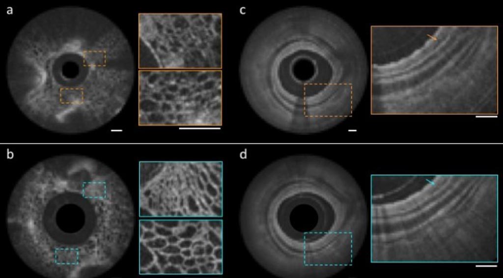
Image: Images of fruit flesh (L) and swine airways (R) obtained with the nano-optic endoscope (b,d) and a conventional catheter (a,c) (Photo courtesy of Harvard University/MGH).
The tailored chromatic dispersion of the metalens--in the context of spectral interferometry--is utilized to maintain high-resolution imaging beyond the input field Rayleigh range, thus easing the trade-off between transverse resolution and depth of focus. To demonstrate the imaging quality of the nano-optic endoscope, the researchers imaged fruit flesh, swine and sheep airways, and resected human lung tissue. The images captured clearly showed cellular structures in fruit flesh and tissue layers and fine glands in the bronchial mucosa of swine and sheep.
In the human lung tissue, the researchers were able to clearly identify structures corresponding to the fine, irregular glands indicating the presence of adenocarcinoma, the most prominent type of lung cancer. The researchers now aim to explore other applications for the nano-optic endoscope, including a polarization-sensitive nano-optic endoscope, which could contrast between tissues that have highly organized structures, such as smooth muscle, collagen, and blood vessels. The study was published on July 30, 2018, in Nature Photonics.
“Metalenses based on flat optics are a game changing new technology, because the control of image distortions necessary for high resolution imaging is straightforward compared to conventional optics, which requires multiple complex shaped lenses,” said co-senior author professor Federico Capasso, PhD, of the Harvard University School of Engineering and Applied Sciences (SEAS). “I am confident that this will lead to a new class of optical systems and instruments with a broad range of applications in many areas of science and technology.”
“Clinical adoption of many cutting-edge endoscopic microscopy modalities has been hampered due to the difficulty of designing miniature catheters that achieve the same image quality as bulky desktop microscopes,” said co-senior author Melissa Suter, MD, of MGH. “The use of nano-optic catheters that incorporate metalenses into their design will likely change the landscape of optical catheter design, resulting in a dramatic increase in the quality, resolution, and functionality of endoscopic microscopy.”
Instead of using solid pieces of curved class to focus light, metalens’ are covered with an array of titanium dioxide nanofins that helps focus light on a point in exactly the same way. A major benefit of the metalens is that it is able to completely eliminate chromatic aberration, a common issue in optical lenses. Chromatic aberration occurs when a lens fails to focus all colors on the same point, causing rainbow edges to appear in areas of contrast in photos.
Related Links:
Massachusetts General Hospital
Harvard University


