Radiation Beam Imaging System Observes RT in Action
By MedImaging International staff writers
Posted on 10 Aug 2018
A novel imaging system provides real-time visualization of radiotherapy (RT) beam shape at the point of incidence and exit.Posted on 10 Aug 2018
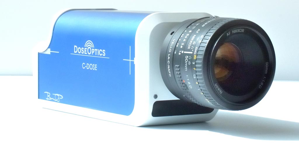
The DoseOptics (Lebanon, NH, USA) C Dose RESEARCH camera uses unique time-gating video technology to capture both photon and electron beam delivery and ensure that each pulse of the linear accelerator contributes to the image, with time-integrating software allowing a cumulative image that can be overlaid in real time on the object being irradiated. Information on RT cone position, gantry movements, and the multi-leaf collimator (MLC) jaw are recorded simultaneously. The camera and software operate remotely, providing an independent check and measurement tool for both beam shape and delivery.

Image: A novel imaging system visualizes RT in real time (Photo courtesy of DoseOptics).
The C Dose RESEARCH camera is based on detection of Cherenkov radiation, an electromagnetic (EM) radiation emitted when a charged particle (such as an electron) passes through a dielectric (phased) medium at a speed that is greater than the phase velocity of light in the same medium. This gives off a visible glow. One such example of Cherenkov radiation is the characteristic blue glow emitted by an underwater nuclear reactor. Cherenkov radiation is named after Soviet scientist Pavel Cherenkov, who won the 1958 Nobel Prize in physics.
“We are extremely excited to be able to offer a Cherenkov imaging system to the field of radiation oncology where we believe it can change the paradigm of radiation delivery verification, and provide intuitive visualization of the treatment for everyone in the department,” said Brian Pogue, MD, PhD, president of DoseOptics. “As new delivery techniques improve and become more and more complex, verification remains a challenge. With C-Dose, medical physicists charged with ensuring delivery accuracy can now literally see what they are doing.”
Current external beam radiation therapy (EBRT) is a “blind” procedure, with patients aligned and imaged via computerized tomography (CT). Treatment plans are generated at the instrument before treatment starts. While EBRT is a highly precise treatment with little room for error, routine verification of targeted RT delivery is not typically done. Major errors are estimated at about 0.2%, and minor errors are likely to occur much more frequently. The consequences can range from skin burns to tissue damage to death.
Related Links:
DoseOptics


