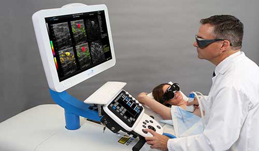
Optoacoustic Imaging Augments Breast Mass Assessment
By MedImaging International staff writers
Posted on 06 Jun 2018
A novel breast imaging system fuses optoacoustic (OA) and ultrasound (US) technologies to generate real-time functional and anatomical images of the breast.Posted on 06 Jun 2018
The Seno Medical Instruments (San Antonio, TX, USA) Imagio OA/US breast imaging system uses OA to provide a “blood map” around breast masses, while US provides a traditional anatomic image. Unlike current techniques that create images by transmitting and receiving energy in the same form, OA/US imaging transmits photonic energy, but detects acoustic energy; by transmitting multiple bandwidths of laser light, a much broader range of data can be captured, which makes the functional images possible.

Image: A novel breast imaging system fuses optoacoustics and ultrasound (Photo courtesy of Seno Medical).
The system can thus detect the appearance (or absence) of two of the hallmark indicators of cancer, angiogenesis and deoxygenation, by measuring hemoglobin concentration in the breast tissues. The resulting amalgamation of data helps radiologists confirm or rule out malignancy with more certainty than current diagnostic imaging modalities, and without exposing patients to potentially harmful ionizing X-ray radiation or contrast agents.
A study by researchers at the Northwestern University (NU; Chicago, IL, USA) and other institutions involving 2,105 women conducted to compare the diagnostic utility of the Imagio OA/US breast imaging system to grayscale US alone, found that OA/US downgraded 40.8% of benign mass reads, with a specificity of 43%, compared to 28.1% for grayscale US alone, resulting in a need for fewer subsequent needle biopsies. The study was published in the May 2018 issue of Radiology.
“Needle biopsies are expensive, very stressful to the patient, often require another appointment for the patient, and it can take days to get the results. Patients who are safely downgraded to not needing a biopsy would benefit,” said Stephen Grobmyer, MD, of the Cleveland Clinic (OH, USA) Comprehensive Breast Cancer Program. “Safely reducing the number of breast biopsy procedures through advanced imaging would add a lot of value to the current system. It also may help identify lesions that require biopsy that were thought to be benign by traditional imaging.”
OA is a biomedical imaging modality based on the photoacoustic effect, which results of some of the delivered energy absorbed and converted into heat, leading to transient thermoelastic expansion and thus wideband US emission. As optical absorption is closely associated with physiological properties, such as hemoglobin concentration and oxygen saturation, the magnitude of the US emission reveals physiologically specific optical absorption contrast, allowing 2D or 3D images of the targeted areas to be formed.


 Guided Devices.jpg)


