Shortwave Infrared Fluorescence May Enhance Biological Imaging
By MedImaging International staff writers
Posted on 17 Apr 2018
A new study shows that indocyanine green (ICG) dye also fluoresces in shortwave infrared (SWIR) wavelengths, resulting in images much clearer than those taken in the near infrared (NIR) band.Posted on 17 Apr 2018
Researchers at the Massachusetts Institute of Technology (MIT, Cambridge, MA, USA), Tufts University (Medford, MA, USA), and Massachusetts General Hospital (MGH; Boston, USA) conducted a study to examine if commercially available NIR dyes possess optical properties that could make them suitable for SWIR, as fluorophores can then offer better contrast and clarity than in shorter wavelengths, due to greatly reduced scattering and improved tissue penetration.

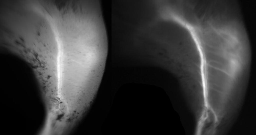
Image: Capillary imaging using NIR (L), as compared to SWIR (R), using ICG dye (Photo courtesy of MIT).
They found that even though their emission spectra peak in the NIR, both ICG and IRDye 800CW dyes outperformed commercial SWIR fluorophores, even beyond 1,500 nm. Real-time fluorescence imaging using ICG at clinically relevant doses, including intravital microscopy, noninvasive imaging in blood and lymph vessels, and imaging of hepatobiliary clearance, demonstrated increased contrast compared with NIR fluorescence imaging. They also found that IRDye 800CW could image cancerous tumors in the SWIR range, by using it to label trastuzumab. The study was published on April 6, 2018, in Proceedings of the National Academy of Sciences (PNAS).
“What we found is that this dye, which has been approved since 1959, is really the best, the brightest fluorophore that we know of at this point for imaging in the short-wave infrared,” said senior author professor of chemistry Moungi Bawendi, PhD, of MIT. “Now clinicians can start to try short-wave imaging for their applications, because they already have a fluorophore which is approved for use in humans.”
High-contrast SWIR fluorescence imaging can be implemented alongside existing imaging modalities by switching the detection of conventional NIR fluorescence systems from silicon-based NIR cameras to new SWIR cameras, which are based on indium gallium arsenide (InGaAs). While these cameras have historically been prohibitively expensive, their prices have been dropping in the past several years.
Related Links:
Massachusetts Institute of Technology
Tufts University
Massachusetts General Hospital


