Isotropic MRI Application Images the Knee in 3D
By MedImaging International staff writers
Posted on 05 Apr 2018
An innovative magnetic resonance imaging (MRI) application can significantly reduce the time required to perform comprehensive diagnostic exams of the knee.Posted on 05 Apr 2018
The Siemens Healthineers (Erlangen, Germany) GOKnee3D app is designed to acquire high-resolution three-dimensional (3D) isotropic images to enable evaluation of the knee in all planes, including double oblique and curved planar. Volume acquisition is based on the controlled aliasing in parallel imaging results in higher acceleration (CAIPIRINHA) SPACE protocol, a proprietary parallel imaging technique used primarily for breath-hold abdominal imaging which enables higher scan speeds and optimal image reconstruction, with better signal quality.
.")
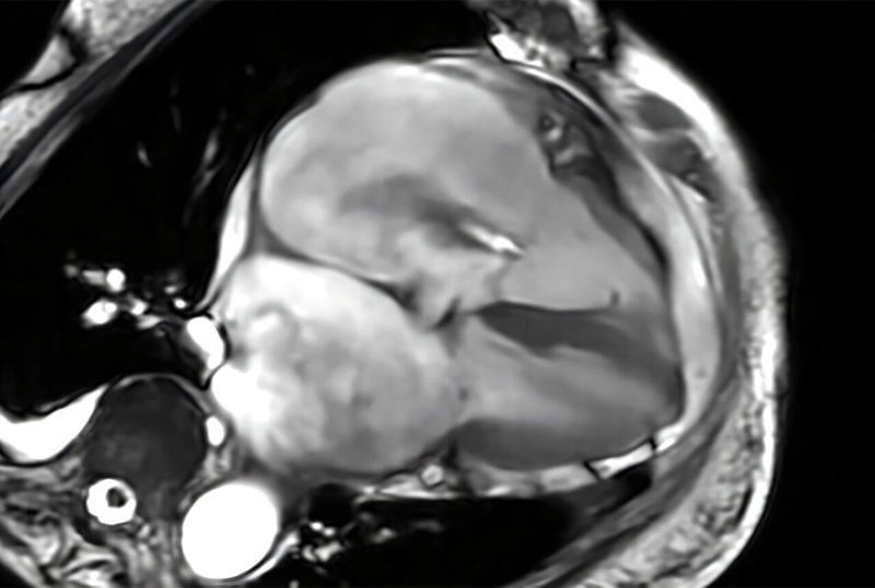
Image: High-resolution 3D isotropic images using the GOKnee3D app (Photo courtesy of Siemens Healthineers).
Supported by dedicated, high-channel Tim 4G knee coils, as well as automated field-of-view adaptation based on machine learning and artificial intelligence (AI), the MR scanner acquires the volume data of the knee joint at the touch of a button, enabling diagnostic 3D knee exam in just 10 minutes. GOKnee3D is available for the Siemens Healthineers Magnetom Aera 1.5T and the Magnetom Skyra 3T MRI scanners, with an eventual rollout planned for additional scanners in the company’s MRI portfolio.
“The fully automated CAIPIRINHA SPACE protocol provides high-quality MR imaging in ten minutes and ensures consistency of image quality and operational efficiency,” said Jan Fritz, MD, assistant professor of radiology and radiological sciences at Johns Hopkins University School of Medicine (Baltimore, MD, USA). “The high spatial resolution isotropic data sets help to visualize abnormalities with high accuracy, enable reformations of virtually any imaging plane, and create high-quality 3D-rendered MR images.”
MRI of the knee provides detailed images of structures within the knee joint, including bones, cartilage, tendons, ligaments, muscles and blood vessels, from many angles. The detailed images allow physicians to evaluate knee function and determine the presence of anomalies and defects. Knee examinations are the third most common type of MRI, accounting for 11% of all scans.

Digital Color Doppler Ultrasound System
MS22Plus

Mobile X-Ray System
K4W

Multi-Use Ultrasound Table
Clinton

Medical Radiographic X-Ray Machine
TR30N HF