Perfusion Imaging of Stroke Victims Improves Emergency Treatment
By MedImaging International staff writers
Posted on 06 Feb 2018
Posted on 06 Feb 2018
A new study suggests that advanced brain imaging can identify stroke patients that could benefit from restoring blood flow in an extended treatment window.

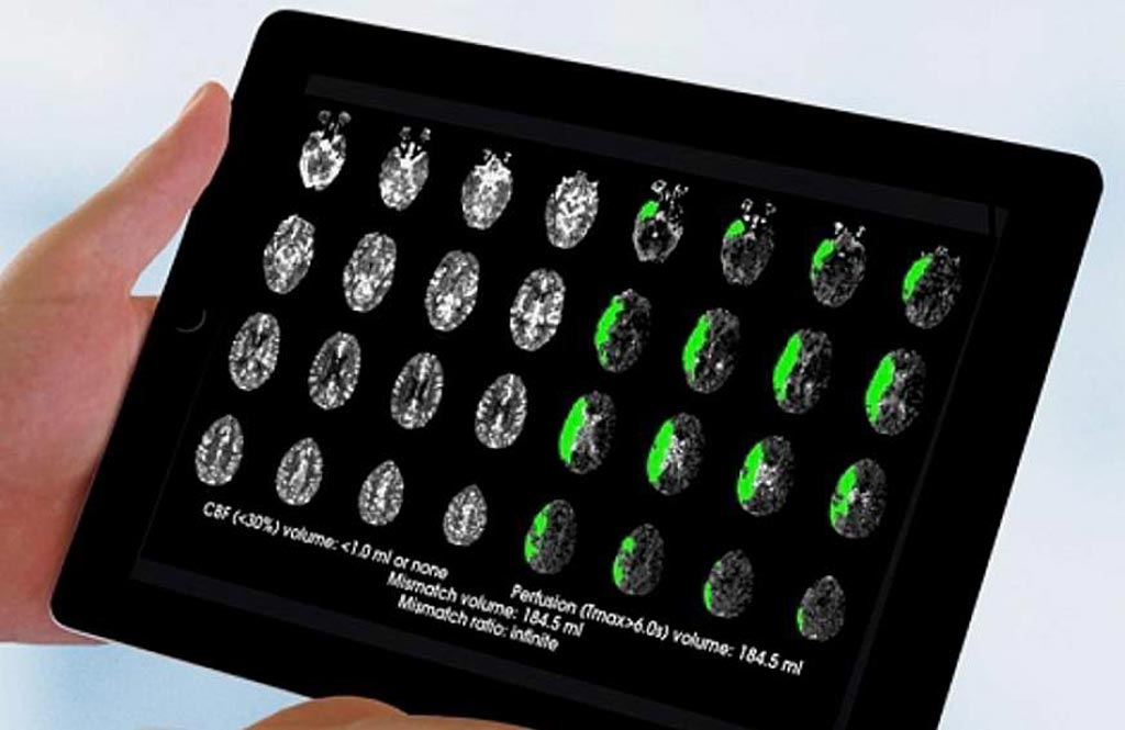
Image: A new study suggests perfusion imaging technology may identify more patients eligible for stroke treatment (Photo courtesy of Greg Albers/ Stanford University).
Researchers at Stanford University School of Medicine (CA, USA), the University of Calgary (Alberta, Canada), the University of Iowa (Iowa City, USA), and other institutions conducted a multicenter, randomized trial of stoke patients who had remaining ischemic brain tissue 6-16 hours after the event, and who had not yet infarcted. All patients underwent perfusion imaging using automated software to analyze perfusion magnetic resonance imaging (pMRI) or computerized tomography (CT) scans to identify patients thought to have salvageable tissue.
In all, 182 patients with proximal middle-cerebral-artery or internal-carotid-artery occlusion were randomly assigned to thrombectomy plus standard medical therapy (92 patients) or standard medical therapy alone (90 patients). The results showed that thrombectomy patients had substantially better outcomes 90 days after treatment than those in the control group. For example, 45% of thrombectomy patients achieved functional independence, compared to 17% in the control group. Thrombectomy was also associated with improved survival, with 14% of the treated group dying within 90 days, compared to 26% in the control group. The study was published on January 24, 2018, in NEJM.
“Endovascular therapy 6 to 16 hours after stroke onset plus standard medical therapy resulted in less disability and a higher rate of functional independence at three months than standard medical therapy alone,” concluded lead author Professor Gregory Albers, MD, of Stanford University. “Studies suggest that infarct growth can occur over a period of several days in patients who do not have reperfusion of ischemic regions. In future trials, later time points could be considered to assess changes in infarct volume.”
“These striking results will have an immediate impact and save people from life-long disability or death,” said Walter Koroshetz, MD, director of the U.S. National Institute of Neurological Disorders and Stroke (NINDS). “I really cannot overstate the size of this effect. The study shows that one out of three stroke patients who present with at-risk brain tissue on their scans improve and some may walk out of the hospital saved from what would otherwise have been a devastating brain injury.”


