MRI Helps Determine 3D Architecture of Human Cervix
By MedImaging International staff writers
Posted on 01 Jan 2018
A new study describes how three-dimensional (3D) magnetic resonance imaging (MRI) can be used to monitor women for weaknesses in the cervix, helping to prevent miscarriage.Posted on 01 Jan 2018
Researchers at the University of Leeds (United Kingdom) conducted a cross-sectional study, which involved high-resolution diffusion tensor MRI (DT-MRI) ex-vivo measurements of seven cervices obtained at hysterectomy for a benign lesion, using a 9.4-T Bruker nuclear magnetic resonance (NMR) spectrometer. A deterministic algorithm was used to visualize underlying fiber organization in order to determine the microarchitecture of the human cervix, and identify occlusive structures in the region corresponding to the internal cervical os.
.")
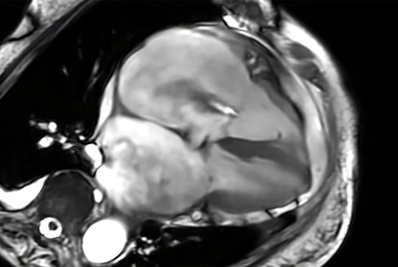
Image: The fibers encircling the cervical canal provide strength and support (Photo courtesy University of Leeds).
The images revealed a fibrous structure running along the upper part of the cervix, which becomes much more pronounced near to where it joins the womb. The fibers are made of collagen and smooth muscle and form a ring around the upper aspect of the cervical canal. During pregnancy, the fibers provide a strong supporting barrier that keeps the fetus and amniotic sac in place, and prevents microorganisms from entering the uterus. During labor, the body releases chemicals that open the cervix, allowing the fetus to enter the birth canal. The study was published on December 11, 2017, in BJOG.
“By applying the imaging techniques that have been used on the brain, we can get a much clearer understanding of the tissue architecture that gives the cervix its unique biomechanical properties,” said Mr. Nigel Simpson, associate professor in obstetrics and gynecology at Leeds University. “This study's findings have encouraged us to explore new imaging techniques to check the integrity of these fibers before or during pregnancy in order to identify at-risk mums, intervene earlier, and so prevent late pregnancy loss and pre-term birth.”
Water molecules undergo random Brownian motion, also known as diffusion. MRI is sensitive to this motion, as controlled by the b-value. When the b-value equals zero, the images are not weighted by diffusion; when the b-value is greater than zero the images are diffusion-weighted. When the diffusion is hindered, by cellular membranes, the myelin shield, etc., the signal is higher. DT-MRI can thus be used to visualize fiber structures, as it can readily differentiate water molecule diffusivities both along and against the fiber.
Related Links:
University of Leeds

Post-Processing Imaging System
DynaCAD Prostate

Digital X-Ray Detector Panel
Acuity G4

Mammo DR Retrofit Solution
DR Retrofit Mammography

Computed Tomography System
Aquilion ONE / INSIGHT Edition