Innovative X-Ray FPS Provides Bedside Imaging Capabilities
By MedImaging International staff writers
Posted on 27 Nov 2017
A new flat-panel source (FPS) makes portable, low-dose, and low-cost three dimensional (3D) digital tomosynthesis (DT) more accessible.Posted on 27 Nov 2017
The Adaptix (Begbroke, United Kingdom) FPS is composed of an array of cold cathode field emitters sealed into a unit, together with a power supply. Each field emitter generates a conelet of X-rays, while a proprietary system, designed to avoid the common problem of high-voltage switching, allows for each X-ray emission to be addressed and individually controlled, thus eliminating large numbers of overlapping X-rays. An additional benefit of the design is that the FPS can cover many different angles, allowing depth information to be derived through DT.

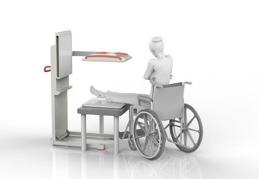
Image: New X-ray panels can generate 3D images at the bedside (Photo courtesy of Adaptix).
The multi-angle imaging is possible without the need to physically move the emitting source, which reduces acquisition time and therefore the risk of motion artifacts. A reduced standoff distance results in reduced power requirements and thermal challenges, compared to conventional X-ray sources, and a further benefit is that beam focal spots are in the sub-millimeter range, providing enhanced resolution. A proprietary image reconstruction solution, developed in conjunction with the University of Oxford (United Kingdom; www.ox.ac.uk), uses sparse data techniques to optimize image reconstruction.
“Avoiding the need to take patients from ICU to the imaging suite for a CT exam would be valued by clinicians and administration in order to avoid the loss of staff on the ward; typically, a transit from ICU to radiology commits a doctor, nurse, and porter for circa 30 minutes,” said the company in a European Commission report summary. “If 3D imaging was available in the ICU it would probably be used more often to answer clinical questions which may be of further value.”
Cold-cathode vacuum tubes do not rely on external heating to provide thermionic emission of electrons. One example of the technology is the Neon lamp, used to produce light as indicators and for special-purpose illumination. The addition of a trigger electrode allows glow discharge to be initiated by an external control circuit. Cold cathodes vacuum tubes sometimes have a rare-earth coating to enhance electron emission.
Related Links:
Adaptix


