MRI Calculates Kidney Scarring without Biopsy
By MedImaging International staff writers
Posted on 15 Sep 2017
Noninvasive magnetic resonance imaging (MRI) elastography can measure kidney damage and predict future kidney function within one year, according to a new study.Posted on 15 Sep 2017
Researchers at St. Michael’s Hospital (Toronto, Canada) conducted a study in 16 patients (mean age 55 years) who underwent MRI elastography following a kidney transplant in order to determine if MRI-based elastography could measure organ stiffness to help estimate fibrosis in the allografts, and predict progression of allograft dysfunction. All participants first underwent free-breathing, flow-compensated elastography on a 3.0-T MRI scanner. They subsequently underwent serial estimated glomerular filtration rate (eGFR) measurement after the elastography scan for a follow-up period of up to one year.
.")
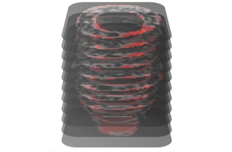
Image: In a new study, MRI elastography demonstrates heterogenous distribution of stiffness in the kidney (Photo courtesy of Anish Kirpalani).
The mean stiffness value of the kidney allograft was compared with both the histopathologic Banff fibrosis score and the rate of eGFR change during the follow-up period. The results revealed that whole-kidney mean stiffness ranged between 3.5 and 7.3 kPa, and correlated with the biopsy-derived Banff fibrosis score. The researchers also found negative correlations between whole-kidney stiffness and both baseline eGFR and eGFR change over time; but irrespective of baseline eGFR, increased kidney stiffness was associated with a greater eGFR decline. The study was published on August 30, 2017, in Clinical Journal of the American Society of Nephrology.
“The MRI allowed us to get a full picture of the kidney, whereas with a biopsy we would only see a tiny piece. We were able to tell that in some parts of the kidney it's very stiff, and in others, it's not stiff at all, which is information we couldn't get from a biopsy,” said lead author radiologist Anish Kirpalani, MD. “Scarring is a big problem for transplant patients, and with MRI we may be able to better guide how kidney transplant patients are treated early on to improve their long-term outcomes.”
“Clinicians are hesitant to send patients for a test that has risks such as internal bleeding, unless a diagnosis can't be made without it. With this new MRI test, doctors can gather valuable information in the many patients for whom the risks of a biopsy are too high,” concluded senior author nephrologist Darren Yuen, MD. “This new MRI test may help facilitate the testing of new anti-scarring treatments. By providing a needle-free way to measure kidney scarring, we may create more opportunities for this crucial research into finding an effective anti-scarring treatment.”
Related Links:
St. Michael’s Hospital

Portable Color Doppler Ultrasound Scanner
DCU10

Portable X-ray Unit
AJEX140H

Ultrasound Table
Women’s Ultrasound EA Table

Digital Radiographic System
OMNERA 300M