MRI Reveals Spinal Atrophy Tied to MS Progression
By MedImaging International staff writers
Posted on 17 May 2017
A new magnetic resonance imaging (MRI) study among early stage multiple sclerosis (MS) patients reveals detectable cord gray matter atrophy in both the cervical and thoracic cord.Posted on 17 May 2017
Researchers at the University of California, San Francisco and University Hospital Basel enrolled 64 patients with early MS and 53 controls in an observational study. The MS patients (mean age 36.9, 26 women) had a mean duration from first symptom onset of 1.2 years, and a median expanded disability status scale (EDSS) score of 2. Among the healthy controls, the mean age was 37.4 and 38 were women.
.")
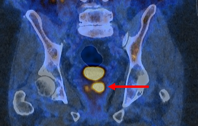
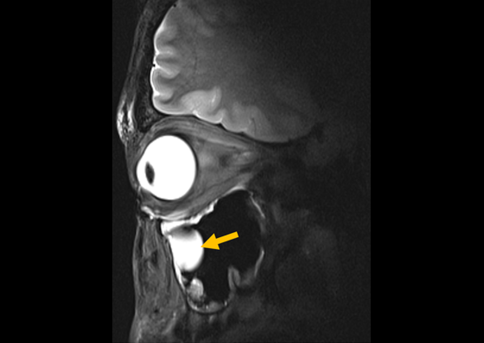
Image: Inversion recovery images illustrating gray matter atrophy in MS (Photo courtesy of Regina Schlaeger).
MRI scans of the C2/C3 cervical spine among the early MS patients revealed a 14% reduction in gray matter area among patients diagnosed with progressive MS, compared with a 3% reduction in gray matter area among patients diagnosed with relapsing MS; the 3% decrease in gray mater area in the cervical spine was also significantly lower than in controls. Total spinal cord area matter was reduced by 10% among patients with progressive disease, but actually increased by a non-significant 2% from controls.
There was also a significant difference in white matter, reduced 9% in those with progressive disease compared with an increase of 3% among patients diagnosed with relapsing MS. The researchers did not find significant differences in the loss of gray matter in the spinal cord at the T9/T10 location among patients with or without spinal cord lesions, but there was a significant difference in spinal cord white matter. In addition, cervical and thoracic gray matter areas were not correlated with the number of spinal cord T2-lesions. The study was presented at the annual American Academy of Neurology meeting, held during April 2017 in Boston (MA, USA).
“Compared to later stages, much of the Expanded Disability Status Score variance is explained by spinal cord lesions at the early stages of disease,” said lead author and study presenter Regina Schlaeger, MD, PhD, of University Hospital Basel. “Spinal cord gray matter atrophy is detectable in vivo already at the earliest stages of multiple sclerosis, affecting both the cervical and lower thoracic cord. In patients without prior spinal cord relapses, cervical cord gray matter area is inversely associated with disability.”
“The presence of lesions at the cervical and lower thoracic spine in the early disease state may be more important in determining a patient's disability, whereas later the amount of gray matter loss may be more relevant in determining a patient's disability,” commented discussant Daniel Reich, MD, PhD, of the U.S. National Institute of Neurological Disorders and Stroke (Bethesda, MD, USA). “All told, I think this is more evidence that early aggressive treatment that can stop lesions from forming, and protect myelin from inflammatory damage, and repair the myelin within existing lesions really will help.”
Historically in neuroscience, the vast majority of research effort has been invested in understanding and studying gray matter and neurons, while white matter has received relatively little attention, largely due to the lack of effective research tools to study white matter, even though it comprises about half the volume of the brain.
 Guided Devices.jpg)
Ultrasound-Guided Biopsy & Visualization Tools
Endoscopic Ultrasound (EUS) Guided Devices

Digital Radiographic System
OMNERA 300M

Ultrasound Table
Women’s Ultrasound EA Table

MRI System
nanoScan MRI 3T/7T