Elastography Imaging Assesses Pituitary Tumor Stiffness
By MedImaging International staff writers
Posted on 17 Feb 2017
A new study reveals that magnetic resonance elastography (MRE) can reliably identify pituitary macroadenoma (PMA) tumors soft enough for removal with minimally invasive surgery.Posted on 17 Feb 2017
Researchers at the Mayo Clinic and the U.S. National Institute of Biomedical Imaging and Bioengineering conducted a study to prospectively evaluate MRE in 10 patients (mean patient age 59.5, 50% male) undergoing trans-sphenoidal resection of a PMA over two centimeters in maximum diameter; the patients were prospectively imaged with MRE. The surgeon, who was blinded to MRE results, graded tumor consistency at surgery as soft, intermediate, or firm.

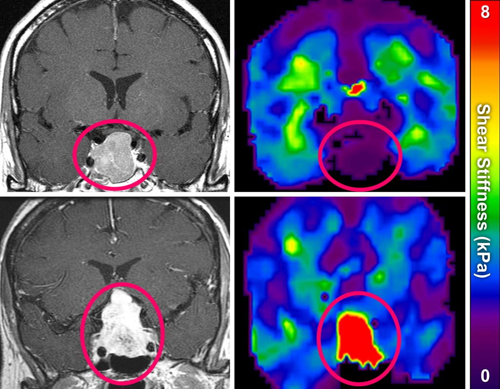
Image: A soft PMA (purple) compared to a hard PMA (red) (Photo courtesy of NIBIB).
The surgeons categorized six tumors as soft and four tumors as medium; no tumors were deemed to be hard. The results showed that the MRE measurements reliably identified the viscoelastic consistency of the tumors that were soft enough for removal with a minimally invasive suction technique, compared to harder tumors that require more invasive surgery. The study was published in the January 2017 issue of Pituitary.
“MRE is similar to a drop of water hitting a still pond to create the ripples that move out in all directions. We generate tiny, harmless ripples, or shear waves, that travel through the brain of the patient,” said senior author professor of radiology John Huston III, MD, of the Mayo Clinic. “Our instruments measure how the ripples change as they move through the brain and those changes give us an extremely accurate measure -- and a color-coded picture -- of the stiffness of the tissue.”
“The group developed brain MRE several years ago and is now successfully applying it to clinical diagnosis and treatment,” commented Guoying Liu, PhD, director of the NIBIB Program in MRI. “This development of a new imaging technique followed by its practical application in surgical planning for better patient outcomes is an outstanding example of one of the main objectives of NIBIB-funded research.”
About 90% of PMAs are soft, with a consistency similar to that of toothpaste. Therefore, without MRE, surgeons would routinely plan for transphenoidal resection, employing very thin instruments that are threaded through the nasal cavity to the pituitary gland at the base of the skull, where suction is used to remove the tumor; but in about 10% of the cases, the surgeon will encounter a hard tumor, which may require a fully invasive craniotomy.


 Guided Devices.jpg)


