PET Imaging Could Assist Lung Cancer Treatment Planning
By MedImaging International staff writers
Posted on 18 Aug 2016
A new method of extracting big data from positron emission tomography (PET) images could help quantify lung tumors caused by a genetic mutation.Posted on 18 Aug 2016
Researchers at Dana-Farber Cancer Institute (Boston, MA, USA), Brigham and Women's Hospital (BWH; Boston, AM, USA), and Harvard Medical School (HMS; Boston, MA, USA) used big data radiomics to extract information from PET images in 348 non-small cell lung cancer (NSCLC) patients. The main outcome was association between radiomic features of tumors and epidermal growth factor receptor (EGFR) and Kristen rat sarcoma viral (KRAS) genetic mutations.

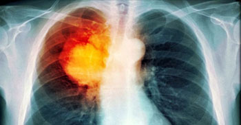
Image: A PET scan image of lung cancer (Photo courtesy of TDMU).
The mutations were confirmed by molecular testing based on biopsies of tumor tissues, the standard of care for mutation identification. The results showed that eight radiomic features describing different aspects of the tumor, such as its shape and textures, appear to be associated with EGFR mutations, but not with KRAS. The researchers suggest that the different metabolic imaging phenotypes quantified by radiomic features may be caused by EGFR mutations, which could guide treatment. For example, 15% of patients with EGFR mutations may benefit from tyrosine kinase inhibitor (TKI) therapies.
The researchers are now assessing combining PET-based radiomic features with features derived from other imaging tests, such as computed tomography (CT) or magnetic resonance imaging (MRI), to improve the accuracy of genetic mutation identification in both lung and brain cancers. The study was presented the 58th annual meeting of the American Association of Physicists in Medicine (AAPM), held during July-August 2016 in Washington (DC, USA).
“Medical images are regularly acquired for every cancer patient that comes into our clinic for treatment, which is the case at many other cancer centers as well,” said lead author Stephen Yip, PhD, an instructor at Harvard Medical School, Dana Farber Cancer Institute and Brigham and Women's Hospital. “This early research suggests that standard-of-care PET imaging may help guide doctors in identifying patients with EGFR mutations, potentially providing valuable information for personalized lung cancer therapy.”
Related Links:
Dana-Farber Cancer Institute
Brigham and Women's Hospital
Harvard Medical School


