Novel Technology Provides Dynamic Respiratory Imaging
By MedImaging International staff writers
Posted on 04 Apr 2016
Innovative Structured light plethysmography (SLP) imaging technology helps construct a 3-dimensional (3D) image that displays regional breathing correlations and lung symmetry. Posted on 04 Apr 2016
The Thora-3Di system projects a structured image that covers the patient’s chest and abdomen to record movement in four dimensions; the recorded image can be replayed in real time, while allowing the clinician to manipulate the view using proprietary SLP software to create a 3D image. The software also provides the operator with the ability to divide the image into defined regions, so as to compare the upper chest and abdomen or the left and right sides of the chest, as a means to identify potential asymmetries indicative of respiratory-related issues.
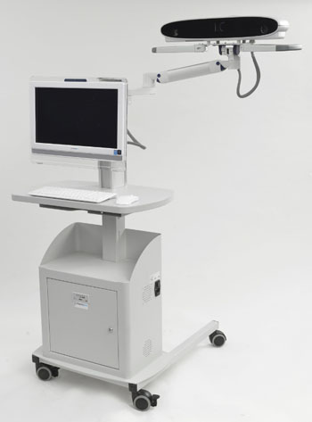
Image: The Thora-3Di system (Photo courtesy of Pneumacare).
By comparing the patient’s symmetry of breathing and measuring the chest wall, Thora-3DI can illustrate the dynamics of lung function in real time, reflecting respiratory function and providing a deeper understanding of the patient's lungs, without the need or forced manoeuver testing or moving the patient to a measurement site. The patient can be sitting, lying, conscious, or unconscious, as the Thora-3DI is noninvasive and does not require any direct interaction with the patient. Thora-3Di is a product of Pneumacare (Cambridgeshire, United Kingdom).
“The measurement of lung function changes in children with respiratory diseases is essential if we are to accurately assess the benefit of treatments. Noninvasive, simple methods have been sought for decades, and the technique developed by Pneumacare is exciting and novel,” said Prof. Warren Lenney, MD, of the University Hospital of North Staffordshire (Stoke on Trent, United Kingdom). “If accurate, reproducible, and sensitive results can be obtained, I can foresee the technique having wide application in all centers specializing in the management of pediatric respiratory illnesses."
Related Links:
Pneumacare




Mammo DR Retrofit Solution
DR Retrofit Mammography


MRI System
nanoScan MRI 3T/7T
Silver Member

X-Ray QA Device
Accu-Gold+ Touch Pro

Ultrasound Table
Women’s Ultrasound EA Table