Imaging Tool Detects Oral Cancer More Accurately
By MedImaging International staff writers
Posted on 15 Nov 2015
A noninvasive device uses fluorescence technology to measure and visualize the biochemical changes that occur in oral epithelial tissue as it turns cancerous.Posted on 15 Nov 2015
Developed by researchers at Texas A&M University (TAMU; College Station, TX, USA), fluorescence lifetime imaging (FLIM) technology noninvasively detects distinct fluorescence signatures specific to benign, precancerous, and cancerous tissue. The system, a small, handheld reflectance confocal laser endomicroscope, detects natural fluorescence emissions from three molecules present in epithelial tissue: collagen, nicotinamide adenine dinucleotide (NADH), and flavin adenine dinucleotide (FAD).

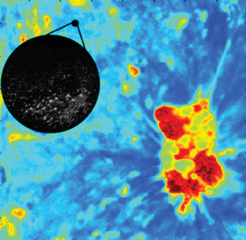
Image: FLIM microscopy detecting oral cancer (Photo courtesy of Texas A&M University).
Collagen has a stronger signal in normal and benign tissue, but in cancerous or precancerous tissue the collagen signals weaken, and NADH and FAD signals increase, since both molecules are related to the energy cells use; malignant cells, per se, use more energy as they rapidly multiply. Another advantage of the system is that that since FLIM detects the natural fluorescence spectrum and lifetime of the molecules, there is no need to use any external contrast agent. Preliminary results from almost 20 patients suggest that FLIM technology can be used to distinguish between benign lesions and dysplasia or squamous cell carcinoma in the human oral cavity.
According to the researchers, FLIM could potentially be used to assist in the clinical management of oral cancer patients, from early screening and diagnosis to treatment and monitoring of recurrence, which happens in 30% of patients who survive a first incidence. Device testing is planned in medical centers in Dallas (TX, USA), Brazil, and Qatar before moving to in-depth clinical trials and commercial licensing. The study presenting FLIM was presented at the World Molecular Imaging Congress, held during September 2015 in Culver City (CA, USA).
“Using FLIM we can see a reduction in the fluorescence lifetime in precancerous tissue and changes in the fluorescence spectrum in malignant tissue that are not apparent in benign tissue,” said study presenter Associate Professor of Biomedical Engineering Javier Jo, PhD. “We then feed this information into a computer where it is processed by an algorithm that essentially color codes the images of the oral cavity; if the tissue is benign it shows up green. If it is cancerous or precancerous, it shows up red.”
The [US] National Institutes of Health (NIH; Bethesda, MD, USA) estimates that more than 8,000 people in the United States will die from oral cancer, and another 37,000 new patients will be diagnosed this year alone. When oral cancer is diagnosed before it spreads, the five-year survival rate is about 80%, but only about 30% of patients are diagnosed at this early stage.
Related Links:
Texas A&M University
US National Institutes of Health


