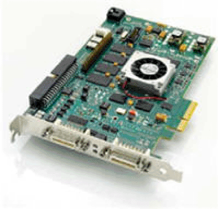
Innovative Digital Image Processor Board Provides Real-Time X-Ray Imaging
By MedImaging International staff writers
Posted on 08 Dec 2009
A digital image processor engineered for demanding X-ray instrumentation and radioscopy is capable of handling a wide range of resolutions, pixel depths, and frame rates.Posted on 08 Dec 2009
The XRI-1600 image processor board is able to generate high quality diagnostic pictures due to the incorporation of a proprietary image processing engine (IPE), specially designed for X-ray imaging applications, which performs real-time digital image processing in three dynamic stages. The first stage is input image conditioning, in which the IPE performs shading correction, lens correction, and image realignment as required; then, a motion-compensated noise reduction algorithm that is optimized for both static and dynamic images is put into effect; and finally an output image conditioning stage that enables image rotation and/or flip, along with image enhancement and masking.

Image: The XRI-1600 Digital Image Processor Board (Photo courtesy of DALSA).
The XRI-1600 is designed for rapid system integration and comes bundled with easy-to-use software application development tools, utilities, and installation scripts to allow rapid application development, diagnosis, and deployment. The XRI-1600 Software Development Kit (SDK) is a Microsoft Windows compatible C++ library for image acquisition and digital image processor control that permit users to control all aspects of the image acquisition process and image storage function, both on the local and host computers. The software features a powerful event notification technology to increase the application response time. Other key features include adaptive image averaging that reduces noise in both still and dynamic images; a programmable digital filter for improving image quality and contrast; a local image storage function that increases reliability and processing time; and flexible input data formats that support high-resolution images.
The XRI-1600 image processor is a product of DALSA (Waterloo, ON, Canada), and was presented at the annual meeting of the Radiological Society of North America (RSNA), held during November-December 2009 in Chicago (IL, USA).
"DALSA's XRI-1600 performs adaptive image processing through the image processing engine to reduce noise in both still and dynamic images and greatly improve image quality and image contrast,” said Inder Kohli, product line manager at DALSA. "This is a key requirement of X-ray and radioscopy applications which typically exhibit low contrast, high-noise, and contain motion artifacts.”
Related Links:
DALSA


