First-Of-Its-Kind Tool Analyzes MRI Scans to Measure Brain Aging
Posted on 25 Feb 2025
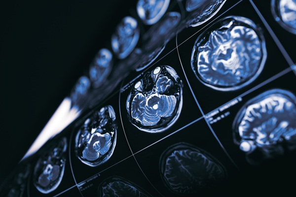
Biological age differs from chronological age, as two individuals of the same age may have distinct biological ages based on the functionality of their bodies and how the tissues appear at a cellular level. Faster brain aging is strongly linked to an increased risk of cognitive decline. Common biological age measures often rely on blood samples to assess epigenetic aging and DNA methylation, which affects gene function in cells. However, using blood samples to gauge biological age is ineffective for determining brain age. The blood-brain barrier prevents blood cells from entering the brain, meaning that blood samples cannot directly reflect aging processes such as DNA methylation in the brain. On the other hand, obtaining a sample directly from the brain is highly invasive, making it impractical to measure DNA methylation and other aging indicators from living human brain cells. Now, researchers have developed a new artificial intelligence (AI) model capable of non-invasively measuring the speed of brain aging, providing an invaluable tool for understanding and addressing cognitive decline and dementia. This innovative model analyzes magnetic resonance imaging (MRI) scans to track brain changes.
Earlier research at the USC Leonard Davis School of Gerontology (Los Angeles, CA, USA; www.gero.usc.edu) revealed the potential of MRI scans to estimate brain biological age without invasive methods. In earlier studies, AI was used to compare a patient’s brain structure with data from MRI scans of thousands of individuals, assessing various ages and cognitive health outcomes. However, the previous model had limitations, as it relied on a single MRI scan to estimate brain age. Although it could indicate whether a patient’s brain was older than their chronological age, it could not determine when the accelerated aging occurred or whether the brain aging process was speeding up.
")
The researchers have developed a new three-dimensional convolutional neural network (3D-CNN) that offers a more accurate method to measure brain aging over time. Trained and validated on more than 3,000 MRI scans from cognitively normal adults, the model's predictive ability was detailed in a study published in the Proceedings of the National Academy of Sciences. Unlike traditional methods that rely on one-time scans, this new longitudinal approach compares baseline and follow-up MRI scans of the same person. This allows for a more precise identification of neuroanatomic changes associated with faster or slower brain aging. The 3D-CNN also produces “saliency maps,” which highlight the specific brain regions that most significantly influence aging speed.
When applied to 104 cognitively healthy adults and 140 Alzheimer’s disease patients, the model’s assessment of brain aging speed showed a close correlation with changes in cognitive function tests at both time points. The researchers suggest that this model could be instrumental in better understanding the trajectories of both healthy aging and neurodegenerative diseases. Moreover, its predictive capabilities could help identify the most effective treatments tailored to individual characteristics. The new model was also able to differentiate the rates of aging in various brain regions, providing insight into how genetic, environmental, and lifestyle factors may influence brain pathology.
The study also noted differences in brain aging between the sexes, which may help explain why men and women face different risks for neurodegenerative diseases, such as Alzheimer’s. The researchers are particularly excited about the potential of this new model to identify individuals experiencing accelerated brain aging before they exhibit any cognitive symptoms. With the introduction of new Alzheimer’s treatments, their effectiveness has been less than expected, possibly because patients have not begun treatment until significant brain pathology has already developed.
“This is a novel measurement that could change the way we track brain health both in the research lab and in the clinic,” said Andrei Irimia, associate professor of gerontology, biomedical engineering, quantitative & computational biology and neuroscience at the USC Leonard Davis School of Gerontology. “Knowing how fast one’s brain is aging can be powerful.”
Related Links:
USC Leonard Davis School of Gerontology >>> www.gero.usc.edu