Enhanced Treatment Assessment for Liver Cancer Using New Imaging Analysis Technique
By MedImaging International staff writers
Posted on 10 Jan 2016
A study presented at the annual Radiological Society of North America (RSNA 2015) meeting in Chicago USA has shown that a novel MRI analysis technique can significantly speed up the assessment of the effectiveness of liver cancer treatment compared to existing methods.Posted on 10 Jan 2016
Hepatocellular Carcinoma (HCC) is the second most deadly cancer worldwide and treatment consists of an image-guided procedure called Transarterial Chemoembolization (TACE). During the procedure chemotherapeutic drugs are delivered to the tumor while at the same time the blood supply to the tumor is blocked. If a patient does not respond to TACE treatment the clinician needs treat them again, or change their therapy, as rapidly as possible. Infiltrative HCC is very difficult to treat after TACE with traditional methods because of the large number of lesions and their ill-defined borders.

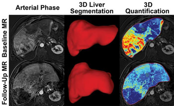
Image: Liver images from before, and after treatment. The bottom-right image shows that less cancer is visible after treatment (Photo courtesy of RSNA).
The researchers used a new approach developed together with Philips Research North America (Cambridge, MA, USA), called the quantitative European Association for the Study of the Liver (qEASL) technique. The new 3D technology provides whole liver volumetric enhancement quantification on Magnetic Resonance Imaging (MRI) and enables a radiologist to segment and delineate an entire tumor in 15–20 seconds in a semi-automated process. The researchers assessed 68 liver cancer patients with infiltrative HCC, using the qEASL technique, before their first TACE procedure, and again one month after the procedure. The researchers measured treatment response, and predicted survival, and segmented the entire liver of the patients while identifying tumors. The researchers found that responders had an overall survival rate of around 21 months and a mean 57.8% decrease in enhancing volume. Non-responders had a survival rate of 6.8 months and a 19.1% increase enhancing volume on average.
According to the researchers, the qEASL approach can also be used with modalities such as cone-beam Computed Tomography (CT), Multidetector CT (MDCT), and Single-Photon Emission Computed Tomography (SPECT), and has also been validated for benign brain, and uterine lesions, and might also be applicable for systemic therapy.
Coauthor of the study, Julius Chapiro, MD, Yale University School of Medicine, said, "In clinical oncology, it is very challenging to assess tumor response to treatment. Up until now, we could measure the extent of tumor diameter or uptake with manual tools like the caliper on the screen, which are highly unreliable due to reader bias. The radiologist can segment the entire tumor with the assistance of the computer. It's a work-flow efficient, semi-automated process that takes 15 to 20 seconds to segment and allows you to delineate the tumor in 3D. The findings show that quantitative tumor enhancement is possible with 3-D qEASL and can predict survival after TACE for infiltrative and multifocal HCC. qEASL is not a diagnostic tool but rather a means of comparing differences before and after treatment to identify non-responders. The earlier the non-responders are identified and treated, the better their outcomes."
Related Links:
Philips Research North America


