AI Tool Measures Lung Nodules with High Accuracy
By MedImaging International staff writers
Posted on 12 Apr 2021
An innovative lung nodule measurement tool built with artificial intelligence (AI) can help accelerate diagnosis of certain respiratory diseases.Posted on 12 Apr 2021
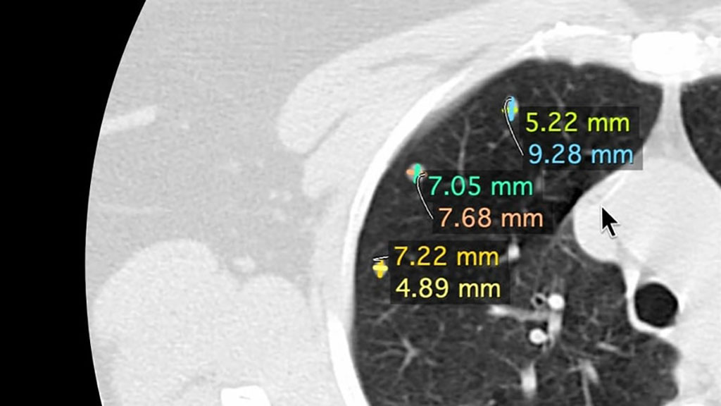
The Nines (Palo Alto, CA, USA) NinesMeasure semi-automatic tool is intended for use by trained radiologists to aid in the analysis and review of adult thoracic computerized tomography (CT) images. NinesMeasure provides quantitative information on pulmonary nodule size on a single study by providing both long and short axis diameter measurements in the axial plane. By doing so, it automates a time consuming, tedious process, as each nodule has to be measured carefully to determine changes in size over time.

Image: NineMeasures automaticall measure a lung nodules axes (Photo courtesy of Nines)
Based on the analysis of digital imaging and communications in medicine (DICOM) data and input from a radiologist that indicates the location of the pulmonary nodule, the device uses AI algorithms to perform the measurements automatically. It can also be used monitor lung nodule size progression and address inter-study consistency spanning a patient's full treatment program. NinesMeasure is strictly limited to analysis of imaging data; in does not replace patient evaluation, nor should it be relied upon to make or confirm a diagnosis.
“In general, radiology is tech-forward in its use of digital imaging, but innovation can make it better,” said David Stavens, PhD, co-founder and CEO of Nines. “Nines has been leading the way by pairing two seemingly disparate groups, skilled radiologists and brilliant engineers, to transform the practice of radiology to be more accessible and more efficient, delivering faster results for quality patient care. That is worth innovating.”
Current lung nodule classification relies on nodule size, a factor that is of limited use for sub-centimeter nodules, or on volume doubling time, a variable that requires follow-up CT exams. As a result, very small lung nodules, with solid components of less than 8 mm in diameter, and therefore below the Lung-RADS 4A risk-stratification threshold, are very difficult to classify, and they are often given a "wait and see" management plan.
Related Links:
Nines


