AI System Improves Accuracy of Cardiac MRI Interpretation
Posted on 26 May 2026
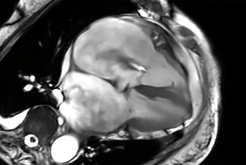
Cardiac magnetic resonance imaging (MRI) is the reference standard for assessing cardiac structure, function, and tissue health. Yet each exam can contain hundreds to thousands of images across multiple views and time points, making interpretation time intensive even for specialists. Demand for cardiac MRI is growing faster than the supply of expert readers, which can delay reporting and access. To help address this challenge, researchers have developed an artificial intelligence system to automate and standardize cardiac MRI interpretation with enhanced accuracy.
The system, called CMR-CLIP, was created by Carnegie Mellon University (Pittsburgh, PA, USA) in collaboration with Cleveland Clinic’s Cardiovascular Innovation Research Center (Cleveland, OH, USA). It connects time-resolved cardiac MRI “videos” with paired clinical radiology reports to learn how physicians describe key findings in practice. By aligning imaging sequences with the report’s impression section, the model learns directly from routine clinical data instead of relying on manually labeled datasets.
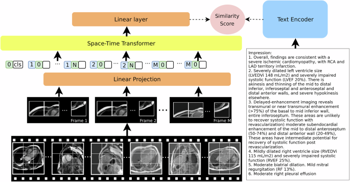
CMR-CLIP represents each study as a set of moving images across standard cardiac views. It processes structure and motion together to mirror how clinicians review a scan. The model was trained on more than 13,000 de-identified Cleveland Clinic patient studies spanning over a decade, incorporating more than a million images and hundreds of thousands of motion sequences. In testing, it identified cardiac conditions in a “zero-shot” setting, linking images to descriptive prompts without prior task-specific labels.
Across benchmarks, the system outperformed general-purpose AI models by up to 35%. With a single example of a condition, it often matched the performance of systems requiring dozens of labeled cases. In specialized diagnostic tasks, it reached near-clinical performance, including accuracies up to 99% for certain conditions. It also retrieved similar cases from large archives using natural-language queries. CMR-CLIP generalized to two external datasets, including one from France and one from Cleveland Clinic Florida, indicating performance beyond the training site.
The team plans to extend CMR-CLIP to perfusion imaging, T2-weighted imaging, and parametric mapping, and to explore automated report generation and interactive decision support. The research was published in Nature Communications, and the model’s codebase is publicly available.
“This work demonstrates that domain-specific foundation models can significantly outperform general-purpose AI systems in specialized clinical applications,” said Ding Zhao, associate professor in Carnegie Mellon University’s Department of Mechanical Engineering and co-principal investigator on the study. “By designing models that reflect the structure and complexity of cardiac MRI data, rather than adapting generic image models, we can unlock new levels of performance and clinical utility.”
“Cardiac MRI interpretation is highly specialized and time intensive. Systems like CMR-CLIP have the potential to support clinicians through automated screening, and interpretation support, particularly in settings where expert readers are limited. Such reader assistant tools are critical to improving patient access to this powerful diagnostic technology,” said David Chen, Ph.D., of Cleveland Clinic and co-principal investigator on the project
Related Links
Carnegie Mellon University
Cleveland Clinic



 Guided Devices.jpg)