Advanced MRI Visualizes Pulsatile Brain Motion
By MedImaging International staff writers
Posted on 18 May 2021
An advanced magnetic resonance imaging (aMRI) technique can capture three dimensional (3D) brain movements in real time, according to a new study. Posted on 18 May 2021
Developed at Mātai Medical Research Institute (Mātai; Gisborne, New Zealand), Stanford University (CA, USA), and other institutions, 3D aMRI builds on previous 2D aMRI, and can show minute movements of the brain at a spatial resolution of just 1.2mm3, about the width of a human hair. The actual movements are amplified by up to a factor of 25 to allow detailed inspection of the movements. The detailed, animated, and magnified movements can help clinicians identify abnormalities, such as those caused by blockages of spinal fluids.

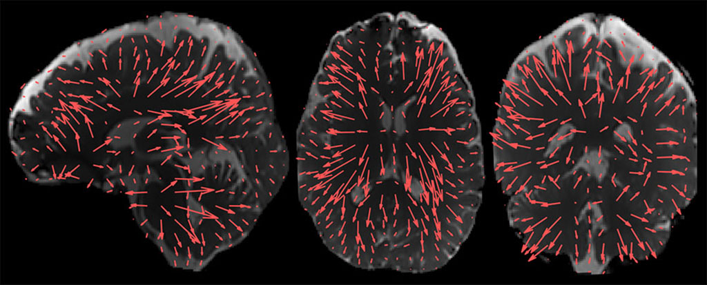
Image: Brain displacement patterns enabled by extra processing of 3D aMRI (Photo courtesy of Mātai)
The researchers also qualitatively compared 3D aMRI to 2D aMRI on multi‐slice and volumetric balanced steady‐state free precession cine data and phase contrast MRI (PC‐MRI), using 3T scans acquired from healthy volunteers. The volumetric 3D aMRI data was also used to generate optical flow maps and 4D animations that reveal complete brain tissue and cerebrospinal fluid (CSF) motion, serving to highlight the piston‐like motion of the four brain ventricles. The study was published on May 5, 2021, in Magnetic Resonance in Medicine.
“The new method magnifies microscopic rhythmic pulsations of the brain as the heart beats to allow the visualization of minute piston-like movements, that are less than the width of a human hair,” said lead author Itamar Terem, MSc, a graduate student at Stanford. “The new 3D version provides a larger magnification factor, which gives us better visibility of brain motion, and better accuracy.”
“This fascinating new visualization method could help us understand what drives the flow of fluid in and around the brain. It will allow us to develop new models of how the brain functions that will guide us in how to maintain brain health and restore it in disease or disorder,” said study co-author Miriam Sadeng, MD, PhD, of the University of Auckland department of anatomy and medical imaging. “The optical flow maps and 4D animations of 3D aMRI may open up exciting applications for neurological diseases that affect the biomechanics of the brain and brain fluids.”
aMRI is based on the use of a phase‐based motion magnification algorithm applied to 2D multi‐slice cardiac gated (cine MRI) data, which results in an amplified “movie” of brain motion. aMRI has also shown promise for visualizing cerebrovascular motion using amplified flow imaging (aFlow), which can capture the characteristics of transient events present in brain tissue, such as blood flow interaction with arterial walls, when applied to 3D PC‐MRA.
Related Links:
Mātai Medical Research Institute
Stanford University


