CT-Based Deep Learning Algorithm Accurately Differentiates Benign From Malignant Vertebral Fractures
Posted on 04 Apr 2024
The rise in the aging population is expected to result in a corresponding increase in the prevalence of vertebral fractures which can cause back pain or neurologic compromise, leading to impaired function or disability. Clinically, benign and malignant vertebral fractures are not distinguishable because they typically occur without adequate trauma. CT imaging plays a key role in distinguishing between benign and malignant vertebral fractures due to the technology’s widespread availability and ability to depict fracture lines on different reconstructed planes. However, distinguishing between benign and malignant vertebral fractures remains challenging with CT alone. Now, a new study has shown that CT-based deep learning models can effectively discriminate benign from malignant vertebral fractures. The study found that the models performed better than or similar to radiology residents and as good as a fellowship-trained radiologist.
In the study, researchers at the Technical University of Munich (TUM, Munich, Germany) examined if the CT-based deep learning models could reliably differentiate between benign and malignant vertebral fractures. The study retrospectively identified CT scans acquired between June 2005 and December 2022 of patients with benign or malignant vertebral fractures based on a composite reference standard that included histopathologic and radiologic information. The researchers randomly selected an internal test set and obtained an external test set from another hospital.
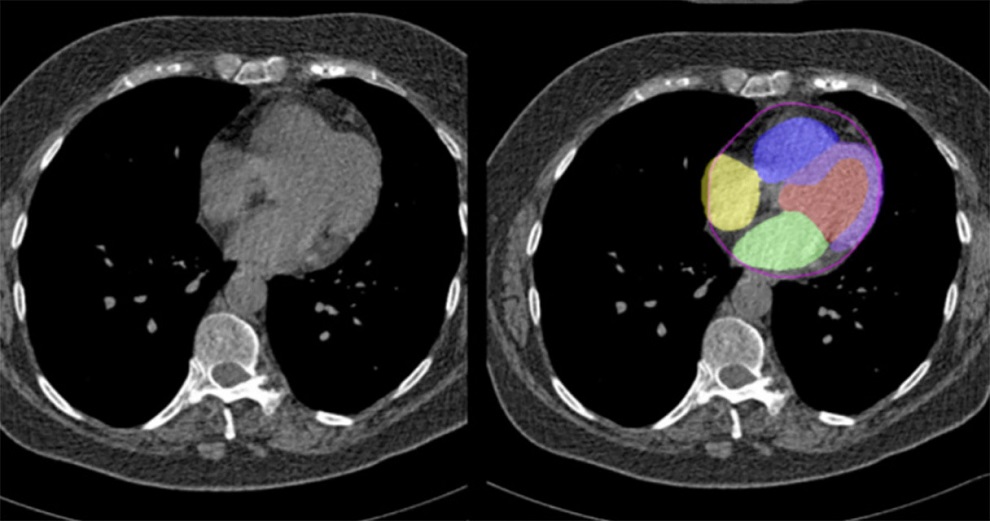
.jpg "Image: Exemplary illustration of the labeling and segmentation process (Photo courtesy of TUM)")
The CT-based deep learning models utilized three-dimensional U-Net encoder-classifier architecture and applied data augmentation during training. The researchers evaluated the models’ performance using the area under the receiver operating characteristic curve (AUC) and compared it with that of two residents and one fellowship-trained radiologist using the DeLong test. The study revealed that the developed models had high discriminatory power for differentiating between benign and malignant vertebral fractures. Their performance surpassed or equaled that of radiology residents and matched that of a fellowship-trained radiologist.
Related Links:
TUM