PET Scans Reveal Hidden Inflammation in Multiple Sclerosis Patients
Posted on 30 Apr 2024
A key challenge for clinicians treating patients with multiple sclerosis (MS) is that after a certain amount of time, they continue to worsen even though their MRIs show no change. A new study has now revealed that advanced brain imaging can detect hidden inflammation in MS patients which traditional MRIs fail to pick up, potentially leading to improved treatment strategies.
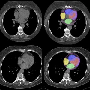
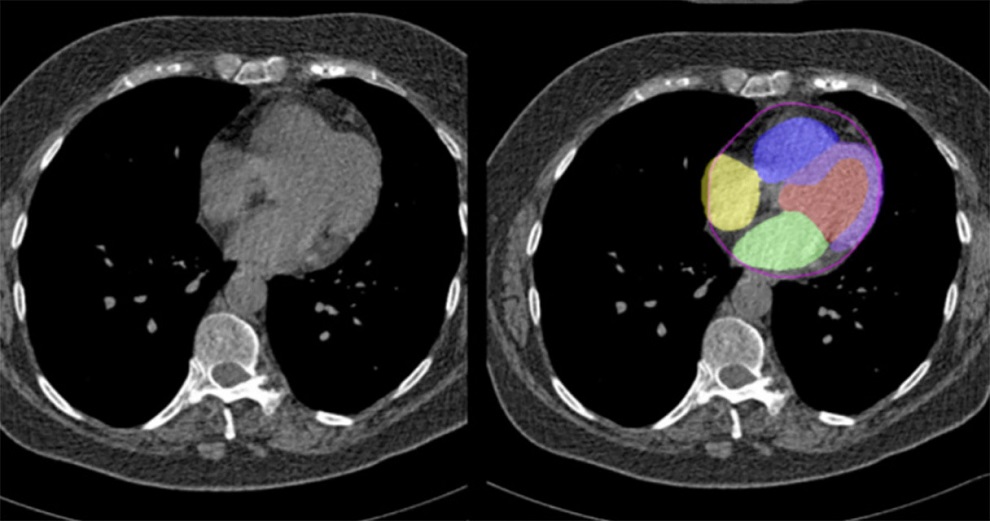
Researchers from Brigham and Women’s Hospital (Boston, MA, USA) have discovered that positron emission tomography (PET) brain scans can identify hidden inflammation in patients undergoing treatment with highly effective MS therapies. This finding emerged when the research team noted that patients on the most effective MS treatments still experienced symptom progression. Over the past eight years, they have developed a technique to image microglia, immune cells in the brain involved in MS progression, which are not visible on routine MRIs. They utilized F18 PBR 06 PET imaging, which uses a tracer binding to microglia cells, revealing that increased microglial activity is associated with greater atrophy of gray matter in the brain, impacting cognition, movement, and fine motor skills.
")
The researchers have coined the term “smoldering” inflammation, comparing it to a smoldering fire that burns slowly without emitting any smoke or flame but continues to cause damage despite showing no signs on MRI. They conducted PET scans on 22 MS patients and eight healthy controls, measuring glial activity load, a new measure developed by the researchers to assess the level of smoldering inflammation from microglia. They found that PET scans showed the hidden inflammation caused by microglia missed by MRI, and the damage to patients’ brains correlated with the patients' levels of disability and fatigue.
Furthermore, the study allowed the researchers to successfully differentiate between patients on high-efficacy and low-efficacy MS treatments. Those on low-efficacy treatments exhibited more PET scan abnormalities, indicating increased microglial activation. Conversely, patients on high-efficacy treatments showed fewer abnormalities but still displayed increased microglial activity compared to healthy individuals. This suggests that while high-efficacy treatments reduce neuroinflammation, residual inflammation persists and may contribute to the worsening of symptoms known as progression independent of relapse activity (PIRA). This research underscores the significant potential of PET scanning in detecting microglial activation, offering critical insights into managing MS more effectively.
“Our therapies are excellent in that we’ve definitely improved MS patients’ lives,” said Rohit Bakshi, MD, of the Department of Neurology. “There’s no doubt about that, but we’re still not at the finish line. This study tells us something new about the disease and may be giving us an important clue as to what is driving disease progression in patients.”
Related Links:
Brigham and Women’s Hospital