Updated Human Brain Map Reveals Uncharted Regions
By MedImaging International staff writers
Posted on 04 Aug 2016
A sharper, multilayered map of the brain will shed light on how our brains work, and could also prove a boon to neurosurgeons as they decide where to insert their scalpels.Posted on 04 Aug 2016
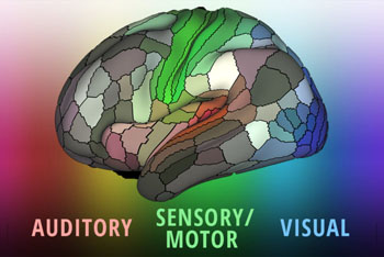
Researchers at Washington University School of Medicine (WUSTL; St. Louis, MO, USA) and Radboud University Medical Centre (Nijmegen, the Netherlands) used multi-modal magnetic resonance imaging (MRI) and functional MRI scans from 210 healthy young adults in the Human Connectome Project (HCP), together with an objective, semi-automated neuroanatomical approach to delineate 360 distinct sections (180 on each hemisphere) in the human brain, which were bounded by sharp changes in cortical architecture, function, connectivity, and/or topography.

Image: The landscape of the cerebral cortex (Photo courtesy of Matthew Glasser/ Eric Young, WUSTL).
The researchers were thus able to characterize 97 new areas and 83 areas previously reported using post-mortem microscopy or other specialized study-specific approaches. To enable the automated delineation and identification of these areas in new HCP subjects, as well as in future studies, they also trained a machine-learning classifier to recognize the multi-modal ‘fingerprint’ of each cortical area. This classifier succeeded in detecting the presence of 96.6% of the cortical areas in new subjects, replicated the group parcellation, and could correctly locate areas in individuals with atypical parcellations. The new brain map was published on July 20, 2016, in Nature.
“We ended up with 180 areas in each hemisphere, but we don’t expect that to be the final number,” said lead author Matthew Glasser, PhD, of WUSTL. “In some cases, we identified a patch of cortex that probably could be subdivided, but we couldn’t confidently draw borders with our current data and techniques. In the future, researchers with better methods will subdivide that area. We focused on borders we are confident will stand the test of time.”
“Each discrete area on the map contains cells with similar structure, function, and connectivity. But these areas differ from each other, just as different countries have well-defined borders and unique cultures,” said senior author neuroscientist David C. Van Essen, PhD, of WUSTL. “We think it will serve the scientific community best if they can dive down and get these maps onto their computer screens and explore as they see fit.”
In 1907, German neurologist Korbinian Brodmann drew some of the first diagrams of the human cortex by hand, based on differences in cellular architecture that he could see under a microscope. For more than a century, scientists have continued using those maps, which depict 52 regions of the brain, as well as those of other neuroanatomists that followed in Brodmann’s footsteps.
Related Links:
Washington University School of Medicine
Radboud University Medical Centre


