New MR Image Guidance Software Provides Direct Tumor Visualization during HDR Prostate Procedures
Posted on 11 Jul 2024
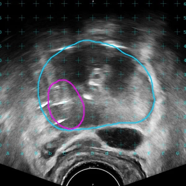
High dose-rate (HDR) brachytherapy is a treatment for prostate cancer that involves placing radioactive sources directly into the prostate gland using needles. For planning such procedures, clinicians traditionally rely on computed tomography (CT) or ultrasound imaging. Integrating magnetic resonance imaging (MRI) into the brachytherapy treatment planning process can minimize toxicity to critical anatomical structures. Nonetheless, difficulties in accurately aligning MRI contours with real-time ultrasound images have limited MRI's application in HDR treatments. Now, an impressive new solution provides visualization of reoriented MRI contours on the live ultrasound during procedures.
GE HealthCare’s (Chicago, IL, USA) MIM Software has introduced MIM Symphony HDR Prostate, a groundbreaking tool designed to enhance HDR brachytherapy. This software aims to boost clinician confidence and enhance patient outcomes by enabling the direct visualization of tumors from MRI scans during live ultrasound-guided HDR prostate treatments. MIM Symphony HDR Prostate facilitates the alignment of MRI-derived contours with real-time ultrasound images, enhancing the visibility of the prostate, any lesions, and critical structures throughout the HDR procedure.

This alignment of the MRI contours with the ultrasound helps clinicians accurately place needles by defining the lesion and tracking changes, thus guiding needle placement accurately as the prostate shifts during the treatment. The software includes ReSlicer, a feature that adjusts for discrepancies between MRI in a supine position and ultrasound in lithotomy positions, and automates needle digitization on CT or ultrasound planning images while also managing needle review and free length checks. MIM Symphony HDR Prostate stands out for its ability to correct MRI orientations and provide MRI-guided visualization during HDR prostate treatments. This new solution is the latest addition to GE HealthCare’s MIM Software portfolio of vendor-neutral solutions for radiation oncology.
“Our goal with this solution is to empower clinicians to plan and deliver safe, more informed, and precise treatment for patients with prostate disease,” said Andrew Nelson, CEO of MIM Software, GE HealthCare. “MIM Symphony HDR Prostate exemplifies decades of MIM Software and GE HealthCare innovations that enable precision care, and we’re thrilled to expand our impact in brachytherapy.”


