Ultrasound Detects Placenta Problems in Small Babies
Posted on 07 Feb 2024
Approximately 10% of fetuses are categorized as small for their gestational age. If these babies are otherwise healthy, there's typically no need for intervention during the pregnancy. However, for small babies experiencing placental dysfunction, proactive measures may be necessary, sometimes including the induction of birth. Therefore, it is crucial to identify which small babies are affected by placental issues. Now, a new study has discovered that Doppler ultrasound, which measures the blood flow in small unborn babies, can indicate the health of the placenta. If deviations are repeatedly observed in these Doppler measurements, further monitoring of the fetus is required. Such deviations signal an increased risk of oxygen deficiency and other health issues for the baby.
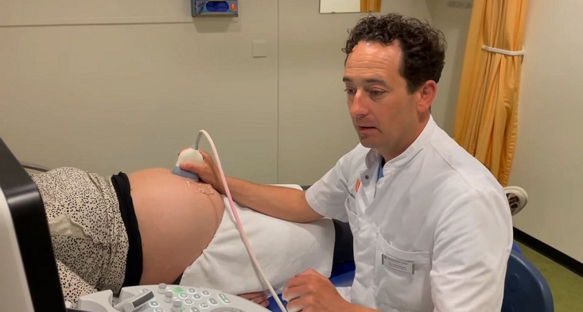
Growth ultrasounds have been utilized for nearly half a century to identify small fetuses and monitor their growth patterns, particularly to see if their growth rate decreases over time. In the study conducted at Amsterdam UMC (Amsterdam, the Netherlands), small babies were given a 'Doppler ultrasound' in addition to standard growth measurements. This ultrasound assesses the resistance in the blood vessels of the umbilical cord, providing insight into the blood flow to the placenta. It can also measure blood supply to the fetus's brain. An unusually high supply can indicate suboptimal placental function. In such cases, the fetus increases blood flow to the brain to mitigate deficiencies caused by the underperforming placenta. Poor placental function elevates the risk of health problems, like oxygen deprivation, and can increase the likelihood of mortality around birth.

The study also investigated whether inducing labor before 37 weeks of gestation led to better outcomes for the child. The findings showed no improvement in outcomes from early induction. Consequently, the recommendation is to delay labor induction until at least 37 weeks of pregnancy, unless additional health risks emerge. This approach is based on the understanding that it is generally better for the baby to remain in the womb as long as possible without additional health risks.
"What was possible with a Doppler ultrasound was already known, but it is not yet standard practice in all hospitals," said Mauritia Marijnen, PhD candidate at Amsterdam UMC and first author of the study. “This research now shows that this measurement certainly has added value for detecting pregnancies in babies that are too small with a malfunctioning placenta.”
"By adding this Doppler ultrasound to the care plan of these undersized babies, the higher risk of problems surrounding childbirth can be better detected and monitored,” added Wessel Ganzevoort, associate professor of obstetrics at Amsterdam UMC and leader of this study. “Small babies for whom the measurement is normal can also be monitored less intensively. There is therefore a greater chance that the delivery will take place naturally, without intervention.”
Related Links:
Amsterdam UMC


