Deltoid Muscle Ultrasound May Help Detect Diabetes
By MedImaging International staff writers
Posted on 24 Jan 2022
A new study suggests that sonographic evaluation of the deltoid muscle could provide a dedicated, simple, and noninvasive method to detect type 2 diabetes (T2D).Posted on 24 Jan 2022
For the study, researchers at Henry Ford Hospital (Detroit, MI, USA) conducted deltoid muscle ultrasound imaging of 124 diabetic patients, who were categorized as obese T2D, non-obese T2D, obese non-T2D diabetes, and non-obese non-T2D diabetes. Three musculoskeletal radiologists (blinded to patient category) measured grayscale pixel intensity of the deltoid muscle and humeral cortex to calculate a muscle/bone ratio for each patient. Age, gender, race, body mass index (BMI), insulin usage, and hemoglobin A1c level were analyzed, and the difference among the four groups was compared.

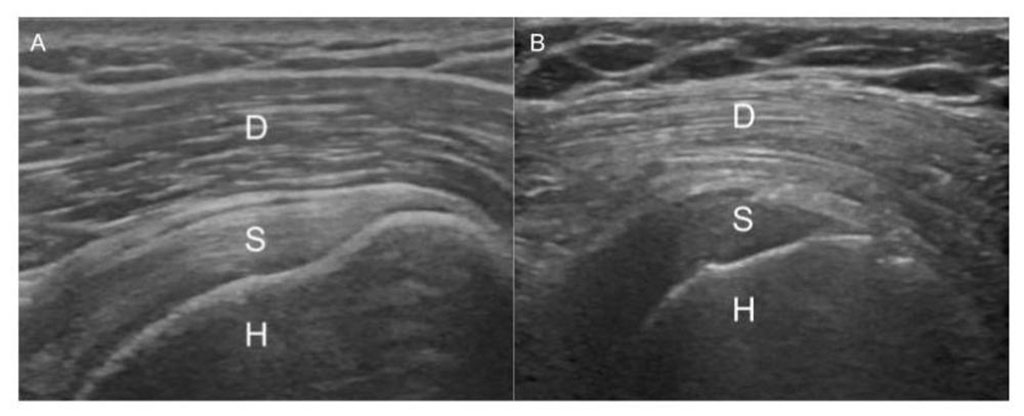
Image: Normal gradient of the deltoid muscle to the supraspinatus tendon (A), and reversal in a T2D patient (D: Deltoid, S: Supraspinatus, H: Humerus) (Photo courtesy of RSNA)
Following baseline measurement, and over a period of three weeks, repeated measurements were done on 40 patients at time. The results showed a statistically significant difference in muscle/bone ratios between the groups; obese T2D - 0.54; non-obese T2D - 0.48; obese non-T2D diabetes - 0.42; and non-obese non-T2D diabetes, 0.35. The overall sensitivity for detecting type 2 diabetes was 80%, with a specificity of 63%. The study was presented at the RSNA annual meeting, held during November 2021 in Chicago (IL, USA).
“Quantitative deltoid muscle ultrasound can detect type two diabetes with the potential for a highly sensitive noninvasive screening method,” concluded lead author and study presenter Steven Bishoy Soliman, DO, RMSK. “This process could translate into a dedicated, simple and noninvasive screening method to detect T2D. The process could help identify some of the 232 million undiagnosed persons globally and could prove especially beneficial in screening of underserved and underrepresented communities.”
In healthy patients, the echogenic appearance of deltoid muscle is darker than that of the underlying rotator cuff tendon. For diabetic patients, the gradient is just the opposite, and the deltoid muscle appears much brighter. The researchers theorized that the brighter appearance is due to low levels of glycogen in the muscle caused by patients’ insulin resistance.
Related Links:
Henry Ford Hospital


