Advanced Ultrasound System Expands Clinical Capabilities
By MedImaging International staff writers
Posted on 30 Apr 2018
An advanced automated breast ultrasound device provides better interpretation of dense breast tissue than traditional mammography.Posted on 30 Apr 2018
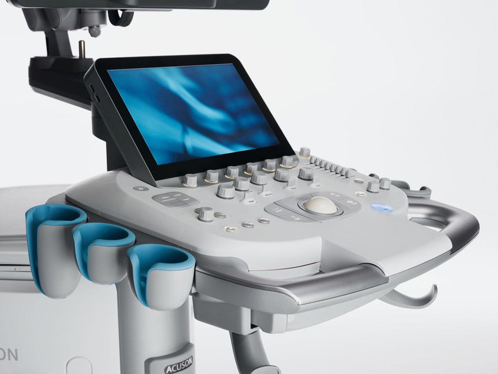
The Siemens Healthineers (Erlangen, Germany) ACUSON S2000 ultrasound system, HELX Evolution with Touch Control is a premium ultrasound system that offers advanced technologies and diagnostic tools to provide sharp, clear images that increase diagnostic confidence. Powered by an intuitive, user-centric interface, the system boasts an intuitive 12.1” high-resolution touch display with instant response technology, a laser-optical trackball, and a simplified home-base control structure that help to optimize exam workflows and reduce training time.

Image: The ACUSON S2000 ultrasound system (Photo courtesy of Siemens Healthineers).
Workflow innovations include context-sensitive body markers, intuitive pictograms, transducer markers, and supportive protocols. Smart-swap annotations with color-coded guidance simplify annotations by suggesting logical replacement text specific to the exam being performed. Innovative ergonomics and digital and tactile keyboards, together with access to proprietary eSieScan workflow protocols increase flexibility for different user preferences and workflow situations, thus allowing fewer motions and inputs, improved operation, and reduced repetitive stress injuries.
System features include HD transducer technology for sharper detail and contrast resolution; full-body coverage; automated breast volume scanning (ABVS) for three dimensional (3D) and hand-held imaging of the breast; tissue strain analytics that provide deeper insights into dense breast tissue; shear wave elastography to evaluate tissue stiffness; in-depth anatomy visualization when using contrast enhanced ultrasound (CEUS); and visualization of lesions in organs using Cadence contrast harmonic imaging (CHI) and Cadence contrast pulse sequencing (CPS) for vascularity and microbubble visualization.
ABVS uses high-frequency sound waves to produce a 3D volumetric image of the entire breast, which allows radiologists to check the breast from multiple angles. Using traditional ultrasound, exams can take up to 30 minutes and are highly impacted by the operator’s skill level due to the device’s handheld transducer. ABVS solves both of these problems by automatically scanning the breast in as little as seven minutes.


