Head Circumference Determines Brain Damage in Newborns
By MedImaging International staff writers
Posted on 19 Mar 2018
A new cranial ultrasound study reveals that a large head circumference at birth increases the risk for brain damage by tenfold.Posted on 19 Mar 2018
Researchers at Ruhr University (Bochum, Germany) and Wilhelmshaven Hospital (WHV; Germany) conducted a study that prospectively screened 4,725 term-born infants by cranial ultrasound in order to explore both the incidence and morphometric risk factors for white matter damage (WMD), an acknowledged prime risk factor for cerebral palsy (CP). The researchers related growth variables and risk factors of the term-born infants using odds ratios of z-score bands.

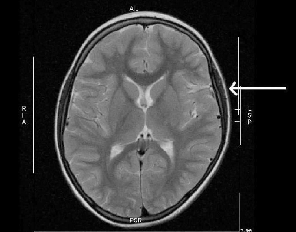
Image: A depression of the brain during birth due to large head circumference (Photo courtesy of Ruhr University).
The results revealed that of the 61 term-born neonates identified with WMD, head circumference was a prime and unique index over the whole range of centiles, suggesting different mechanisms for WMD in the lowest and highest z-score band. While in the highest z-score band, cephalic pressure gradients and prolonged labor with preserved neonatal vitality prevailed, in the lowest band, acute and chronic oxygen deprivation with reduced vitality predominated. The study was published on February 28, 2018, in Obstetrics and Gynecology.
“The fact that seemingly healthy term-born neonates are not screened by head imaging, in spite of both large head circumference and prolonged labor, is considered to be the missing link between the insult that escapes diagnosis and the development of unexplained developmental delay and cerebral palsy in childhood,” said lead author Professor Arne Jensen, MD, of Ruhr-University. “Our findings may elucidate the phenomenon of unexplained cases of developmental delay and CP in the pediatric population on the basis of mechanical constraints and pressure gradients squeezing the skull and brain during parturition when a cephalopelvic disproportion is present.”
Cephalopelvic disproportion exists when the capacity of the pelvis is inadequate to allow the fetus to negotiate the birth canal. This may be due to a small pelvis, a nongynecoid pelvic formation, a large fetus, an unfavorable orientation of the fetus, or a combination of factors. From an evolutionary point of view, upright gait, a growing brain volume, and general growth acceleration will aggravate the problem and will lead to increased caesarean section rates in the highest z-score bands of head circumference in the future to prevent WMD.
Related Links:
Ruhr University
Wilhelmshaven Hospital


