Nanoparticles Demonstrate Use as Injectable Marker
By MedImaging International staff writers
Posted on 01 Aug 2017
Researchers have found a new tissue adhesive that can be detected by clinical imaging modalities, and is also extremely biocompatible for minimally invasive procedures.Posted on 01 Aug 2017
The injectable biocompatible radiopaque Tantalum oxide/Silica core/shell Nanoparticles (TSN) were not only very adhesive, but also demonstrated high-contrast for real-time imaging. Adhesives are becoming a convenient alternative to staples and sutures for reconnecting injured tissues and closing wounds following trauma or surgery, especially in minimally invasive procedures.

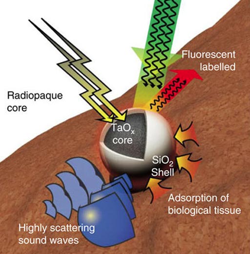
Image: An illustration of a multifunctional Tantalum oxide/silica core/shell nanoparticles (TSNs) with a radiopaque core for X-ray imaging, conjugated fluorescent dye for fluorescent imaging, a dense core material with a high sound-scattering effect for ultrasound imaging, and a silica surface for adhesive property (Photo courtesy of Nature Communications).
The results were published online in the July 19, 2017, issue of the journal Nature Communications by the researchers from the Seoul National University (Seoul, South Korea) and other academic and medical institutions in South Korea. The TSN particles cause significantly less inflammation, and cellular toxicity than CyanoAcrylate and Lipiodol (CA-Lp), imageable tissue adhesives.
TSNs could be used for image-guided procedures such as needle biopsy, catheter-directed embolotherapy, percutaneous angioplasty, vertebroplasty and Radio Frequency (RF) / microwave ablation. These minimally invasive procedures are increasingly used to replacing conventional surgery to control internal bleeding, to perform organ biopsies, and for vessel occlusion for example.
The researchers were able to demonstrate that the TSN glue was clearly visualized by X-Ray fluoroscopy, Computed Tomography (CT), Magnetic Resonance Imaging (MRI), ultrasound, and other clinical modalities. Tantalum oxide is inert, takes time to biodegrade in the body, and is normally only required in very small quantities in image-guided procedures, and remain in the target area without any extreme severe adverse reactions.
Related Links:
Seoul National University


